
Previous issue | Next issue | Archive
![]() Volume 11 (3); September 25, 2021 [Booklet] [EndNote XML for Agris]
Volume 11 (3); September 25, 2021 [Booklet] [EndNote XML for Agris]
 Short Communication
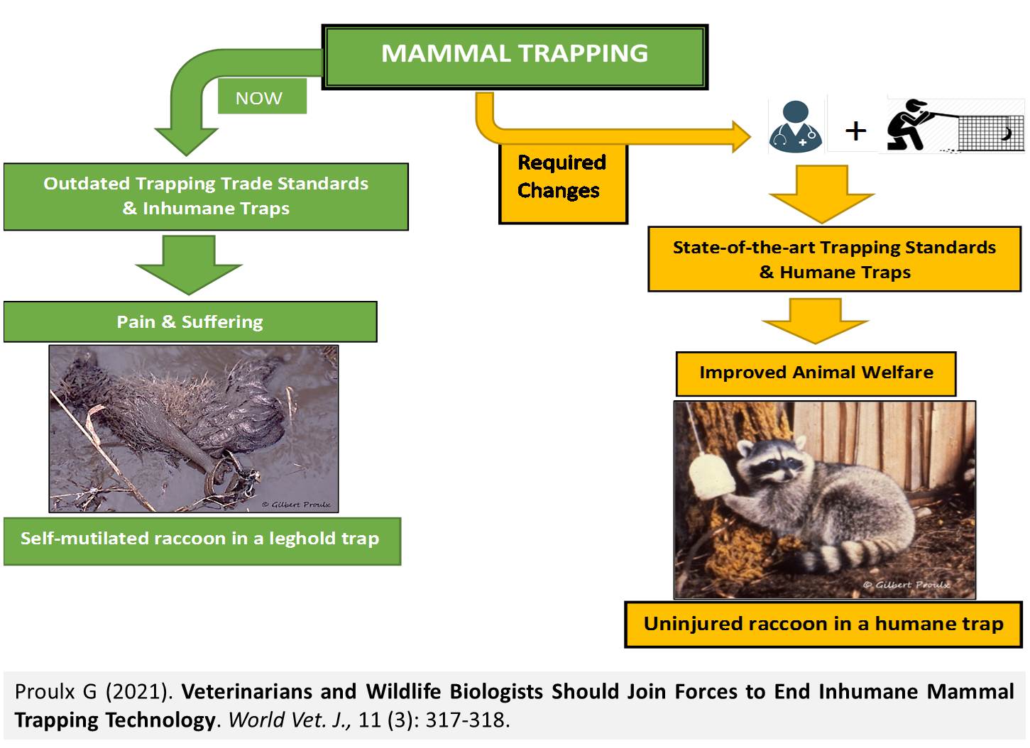
Veterinarians and Wildlife Biologists Should Join Forces to End Inhumane Mammal Trapping Technology
Proulx G.
World Vet. J. 11(3): 317-318, 2021; pii:S232245682100043-11
DOI: https://dx.doi.org/10.54203/scil.2021.wvj43
ABSTRACT: Current mammal trapping standards uphold the use of inhumane trapping technology. For example, killing neck snares for the capture of canids, and rotating-jaw traps, and steel-jawed leghold traps for procyonids and mustelids, are being used by trappers despite decades of research showing that they are inhumane, and cause serious injuries and distress in captured animals. Many wildlife biologists unsuccessfully raised concerns about inhumane mammal trappings. This short communication stresses the need for veterinarians and wildlife biologists to work together to improve the fate of mammals captured in killing or restraining traps, and modify mammal trapping standards on the basis of animal welfare science.
Keywords: Humaneness, Mammal trapping, Traps, Trapping standards
[Full text-PDF] [XML] [Google Scholar] [Crossref Metadata] [Scopus ID: 85115376302] [Export from ePrint] [How to Cite]
 Review
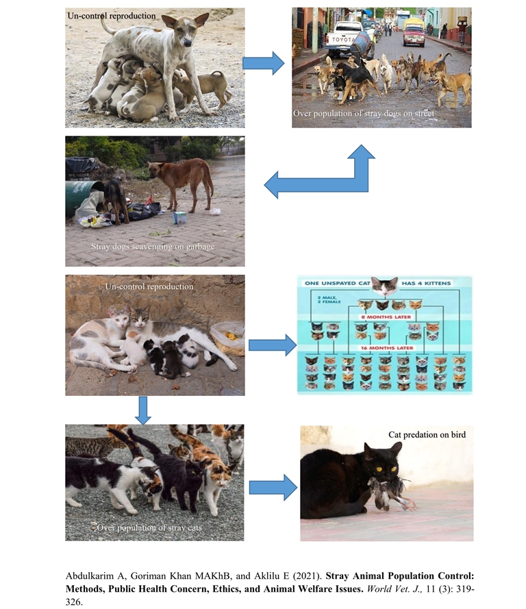
Stray Animal Population Control: Methods, Public Health Concern, Ethics, and Animal Welfare Issues
Abdulkarim A, Goriman Khan MAKhB, and Aklilu E.
World Vet. J. 11(3): 319-326, 2021; pii:S232245682100044-11
DOI: https://dx.doi.org/10.54203/scil.2021.wvj44
ABSTRACT: Stray animal overpopulation has become one of the most serious global problems with many negative impacts on the community, environment, and public health. Most of the stray animals do not depend on humans for food and shelter, and therefore, can reproduce uncontrollably. The uncontrolled reproduction of stray animals increases their population that leads to a higher chance of predation, road traffic accidents, transmission of zoonotic diseases, and therefore, becoming vectors for some diseases. There are several methods for stray animal population control depending on the situation and the nature of the stray animals. These methods include contraceptives, trap-neuter-return, poisoning, euthanasia, and gun shooting among others. Each of the outlined methods has its advantages and disadvantages as to their practicality, ease of conducting, cost, effectiveness, ethics, and animal welfare issues. In conclusion, to achieve successful control measures of the stray animal population and the problem they create, the concerned authorities need to design and enact animal rights laws, provide medical care (treatment and vaccination), feeding, shelter for the animals, and control their reproduction. Public health and environmental agencies may improve the services by regulating personal and environmental hygiene, prevention, and control of zoonotic and transmissible diseases that can be transmitted from stray animals to the public and other livestock respectively.
Keywords: Animal welfare, Ethics, Population control methods, Public health, Stray animals
[Full text-PDF] [XML] [Semantic Scholar] [Crossref Metadata] [Scopus ID: 85115387636] [Export from ePrint] [How to Cite]
 Research Paper
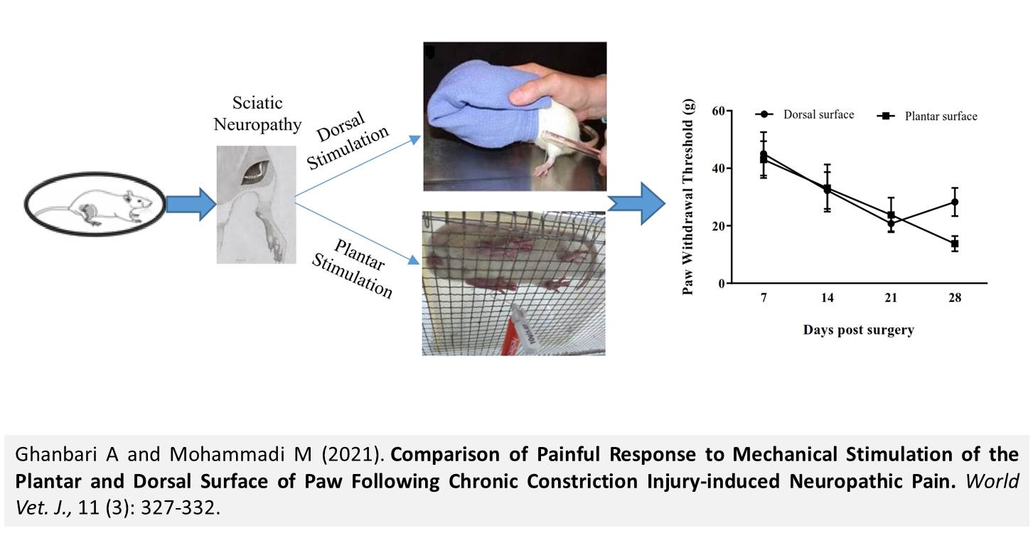
Comparison of Painful Response to Mechanical Stimulation of the Plantar and Dorsal Surface of Paw Following Chronic Constriction Injury-induced Neuropathic Pain
Ghanbari A and Mohammadi M.
World Vet. J. 11(3): 327-332, 2021; pii:S232245682100045-11
DOI: https://dx.doi.org/10.54203/scil.2021.wvj45
ABSTRACT: Mechanical and thermal stimuli were used to evaluate neuropathic pain-like behavior in animal models usually. Mechanical stimulation of paw plantar surface is commonly used to determine mechanical allodynia. In the present study, paw withdrawal response to plantar surface stimulation was compared with paw withdrawal response to dorsal surface stimulation. To this end, a total of 30 female Wistar rats (180-220 g), were assigned randomly to three groups as intact (without any manipulation), sham (incision of skin and muscles without nerve injury), and neuropathy (sciatic nerve lesion) with 10 in each group. To induction of neuropathy (chronic constriction injury), four movable ligations were established around the sciatic nerve using catgut chromic suture with a distance of one millimeter apart and then wound incision was closed. In the sham group, the incision site was closed without nerve ligation. Mechanical allodynia was examined by Von Frey filaments for four weeks. The findings indicated that the paw withdrawal threshold following dorsal surface stimulation was significantly reduced compared to the sham group at day 21 post-surgery. Moreover, paw withdrawal threshold following plantar surface stimulation significantly decreased compared to the sham group at day 21 post-surgery. The present results regarding the sham group showed that the paw withdrawal threshold after mechanical stimulation of the plantar surface was not significantly different from that of the dorsal surface paw. In addition, and there was no significant difference between the paw withdrawal response to plantar surface and dorsal one. In conclusion, paw withdrawal threshold to plantar surface mechanical stimulation was not significantly different from one in dorsal surface following neuropathic pain induced by chronic constriction injury.
Keywords: Mechanical allodynia, Neuropathic pain, Paw dorsal surface, Paw plantar surface, Rat
[Full text-PDF] [XML] [Semantic Scholar] [Crossref Metadata] [Scopus ID: 85115403567] [Export from ePrint] [How to Cite]
 Research Paper
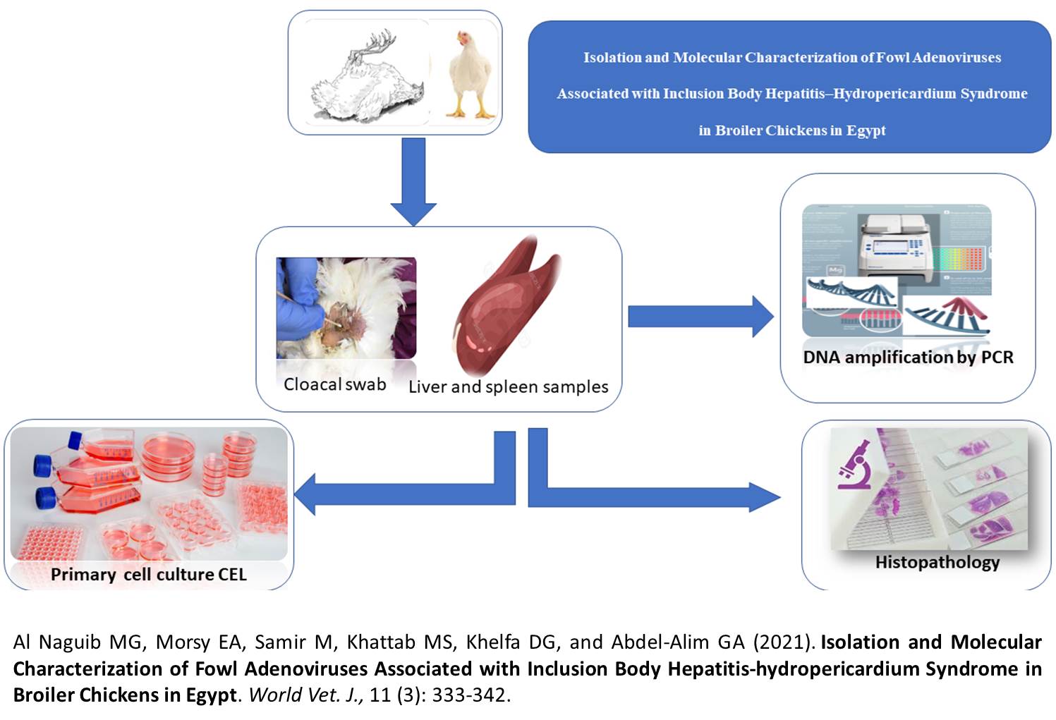
Isolation and Molecular Characterization of Fowl Adenoviruses Associated with Inclusion Body Hepatitis-hydropericardium Syndrome in Broiler Chickens in Egypt
Al Naguib MG, Morsy EA, Samir M, Khattab MS, Khelfa DG, and Abdel-Alim GA.
World Vet. J. 11(3): 333-342, 2021; pii:S232245682100046-11
DOI: https://dx.doi.org/10.54203/scil.2021.wvj46
ABSTRACT: Avian adenoviruses are an extremely diversified group of pathogens that recently triggering a variety of problems for poultry production. In particular, Inclusion Body Hepatitis-Hydropericardium Syndrome (IBH-HPS), which has been observed in broiler birds from 3 to 6 weeks of age and is associated with anemia, hemorrhagic disorders, hydropericardium, and high mortality. The disease has been reported worldwide, and recently it was reported in many Egyptian farms, causing severe economic losses. Therefore, the current study aimed to isolate, and genetically type the most common Adenovirus serotypes associated with this syndrome in Egyptian farms. A total of 50 broiler chicken farms (3-6 weeks old) located in different Egyptian governorates were examined. Macroscopically, the diseased flock revealed hydropericardium, enlarged friable livers with ecchymotic hemorrhages, and varying mortality rates (1 to 7.5%). Histopathologically, severe diffuse necrotizing enteritis, hepatitis, pericarditis, and diffuse lymphoid depletion of the spleen were the most prominent lesions. Liver tissues and cloacal swabs were collected from all examined flocks for FAdVs detection by conventional polymerase chain reaction (PCR) targeting the L1 loop in the hexon gene. The PCR products were sequenced for typing of the detected viruses. It was found that 10 out of 50 flocks examined were PCR positive for FAdVs (20%). Phylogenetic analysis of the sequenced genes revealed that the obtained viruses clustered with reference strains belonging to FAdV type D and E serotype 2, 11, and 8a respectively. The isolation of both FAdV type D and FAdV type E were carried out on a primary cell culture chicken embryo liver cell (CEL) and the presence of these viruses was confirmed by PCR after the appearance of cytopathic effect (CPE). From this study, it could be concluded that both FAdVs types D and E are the most common adenoviruses circulating in poultry farms suffering from hydropericardium and inclusion body hepatitis.
Keywords: Broiler chicken, Chicken embryo liver cell, Fowl adenovirus, Hexon gene, Histopathology, Inclusion body hepatitis–hydropericardium syndrome, PCR
[Full text-PDF] [XML] [Google Scholar] [Crossref Metadata] [Scopus ID: 85115416337] [Export from ePrint] [How to Cite]
 Research Paper
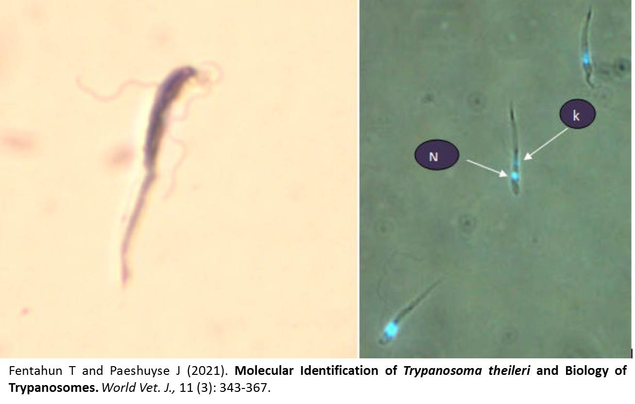
Molecular Identification of Trypanosoma theileri and Biology of Trypanosomes
Fentahun T and Paeshuyse J.
World Vet. J. 11(3): 343-367, 2021; pii:S232245682100047-11
DOI: https://dx.doi.org/10.54203/scil.2021.wvj47
ABSTRACT: Trypanosoma theileri (T. theileri ) is a non-pathogenic, cosmopolitan, and commensal protozoa of cattle. The main objective of the current study was to investigate the biology and feasibility of T. theileri as a model candidate for the discovery of a novel drug. In the present study, the isolates of T. theileri obtained from the Institute of Tropical Medicine (ITM) in SDM 79 were cultivated at 26oC. Eight experiments with different inoculum and different times were grown. The growth curve was plotted to check the growth trends. The doubling time in the logarithmic phase was determined to be 17.43 hours. In addition, an experimental infection was done on a 3-month-old Holstein Friesian calf to isolate the blood-streaming shape; however, it was not successful after the blood buffy coat smear and PBMC culture in RPMI 1640 and HMI 9. Furthermore, the viability was determined by quantitative colorimetric Resazurin assay in 96-well fluorescence Microplates containing 0.4 to 2.4 mM of Resazurin. On the other hand, the response to Pentamidine (1-100 ng/mL) showed a strong negative correlation between the fluorescence signal and the highest Pentamidine concentration. IC50 was 9.25 ng/mL. Genomic DNA was extracted using the phenol-chloroform method. The gradient PCR amplification using T. theileri specific PCR (Tth625-PCR) primers was detected at 465 base pair (bp). In addition, the full-length 18S rDNA sequence was detected at 730 bp. In the silico analysis using common anti-trypanosome drug targets, no significant similarity could be found on either the DNA or the protein level. Nevertheless, homologous sequences have been identified among the drug targets for Ornithine decarboxylase. Therefore, the analysis might show the possibility of using T. theileri as a model for the search of new drugs once they have entire genome sequences. Analysis of the whole genome and transcriptome indicated a phylogenetic relationship between T. theileri and other pathogenic trypanosomes which can be the basis for novel drug development.
Keywords: Drug model, Novel drug, PCR, Resazurin, SDM 79, Trypanosoma theileri
[Full text-PDF] [XML] [Semantic Scholar] [Crossref Metadata] [Scopus ID: 85115358754] [Export from ePrint] [How to Cite]
 Research Paper
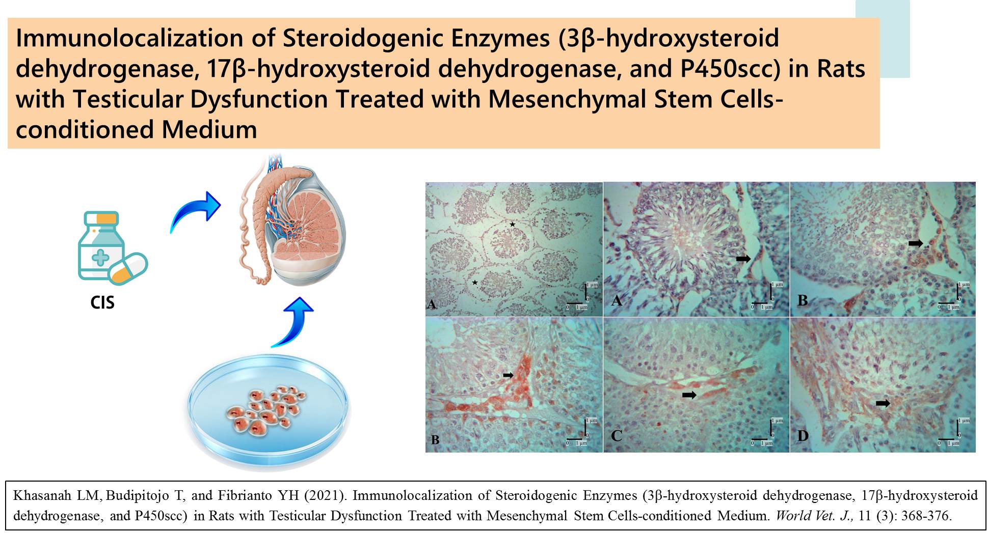
Immunolocalization of Steroidogenic Enzymes (3β-hydroxysteroid dehydrogenase, 17β-hydroxysteroid dehydrogenase, and P450scc) in Rats with Testicular Dysfunction Treated with Mesenchymal Stem Cells-conditioned Medium
Khasanah LM, Budipitojo T, and Fibrianto YH.
World Vet. J. 11(3): 368-376, 2021; pii:S232245682100048-11
DOI: https://dx.doi.org/10.54203/scil.2021.wvj48
ABSTRACT: About 60-80 million couples in the world are suffering from infertility disease. Infertility is a major problem in patients coping with chemotherapy. The chemotherapy process can degenerate non-target organs, especially in testes. Infertility in male or testicular dysfunction is caused by the failure of proliferation and differentiation of the spermatogenic cells. Many studies reported that mesenchymal stem cells-conditioned medium promoted regenerative processes. The present study aimed to investigate the effect of mesenchymal stem cells-conditioned medium on the cisplatin-induced testicular dysfunction by examining the immunolocalization of steroidogenic enzymes, such as 3β-hydroxysteroid dehydrogenase, 17β-hydroxysteroid dehydrogenase, and P450scc which are considered as markers of steroid production. All experimental animals were divided into three groups, namely the control group, mesenchymal stem cells-conditioned medium treated group with an injection dose of 0.2 ml/kg body weight (BW, P1), and mesenchymal stem cells-conditioned medium treated group with an injection dose of 0.5 ml/kg BW (P2). Cisplatin was injected into both treated groups to induce testicular dysfunction. The testicular tissues were processed by the paraffin method, then cut to a thickness of 5 µm, followed by immunohistochemical staining. The HSD3B1 immunoreactivities were found only in Leydig cells, and the intensity increased every week after the injection of mesenchymal stem cells-conditioned medium. The variety of weeks and groups was significantly different in the number of immunoreactive cells of HSD3B1. The results indicated a significant difference between one week after the first injection and the one week after the third and fourth injection. The findings showed a significant difference between the treated group with an injection dose of 0.2 ml/kg BW and the control group. The number of immunoreactive cells of HSD3B1 with an injection dose of 0.5 ml/kg BW was greater compared to the group that received an injection dose of 0.2 ml/kg BW. The intensity of HSD3B1 and HSD17B1 increased every week. The p450scc immunoreactive cells were only found in Leydig cells. The intensity of positive cells of p450scc in the treated group with an injection dose of 0.5 ml/kg BW was more intense, compared to the treated group with an injection dose of 0.2 ml/kg BW. The results of the current study showed that the injection of mesenchymal stem cells-conditioned medium can improve the regeneration of spermatogenic cells, and recover spermatogenesis proved by positive cells of HSD3B1, HSD17B1, and p450scc as markers of steroid production.
Keywords: Cisplatin, HSD17B1, HSD3B1, Mesenchymal stem cells-conditioned medium, P450scc, Testicular dysfunction
[Full text-PDF] [XML] [Google Scholar] [Crossref Metadata] [Scopus ID: 85115431362] [Export from ePrint] [How to Cite]
Download GA as PDF
Research Paper
The Influence of Basil Seed Hydroethanolic Extract on the Skin Wound Healing in Diabetic Male Rats
Hadi M, Moghtadaei-Khorasgani E, and Etesamnia MH.
World Vet. J. 11(3): 377-383, 2021; pii:S232245682100049-11
DOI: https://dx.doi.org/10.54203/scil.2021.wvj49
ABSTRACT: Diabetic wounds and cutaneous wounds are important issues in medical science. Basil is an herbaceous plant and has compounds such as terpenes, flavonoids, and antioxidant properties. A total of 50 male Wistar rats were allocated into 10 groups including the healthy group without treatment, the healthy group treated with 1% phenytoin, eucerin, 5% basil seed extract, 10% basil seed, diabetic group without treatment, 1% phenytoin, eucerin. After anesthesia of rats, we made a 4 cm2 wound on the back of the animal, and different histopathological characteristics were examined, and also on days 3, 7, and 21, the area of wounds was analyzed. In the healthy group treated with 10% basil seed extract, it was revealed that the wound size was significantly healed in the research days. In the diabetic rat groups, the decrease in the wound area was not significant and there was no significant difference between groups. Histopathological findings on day 21 in the healthy group treated with 10% basil seed extract revealed proper epidermis formation and relatively dense dermis containing collagen filaments. While in the diabetic groups, healing was slower. The results indicated that basil extract with anti-inflammatory and antioxidant characteristics can quicken the repair of cutaneous wounds.
Keywords: Basil, Diabetes, Histopathology, Wound
[Full text-PDF] [XML] [Semantic Scholar] [Crossref Metadata] [Scopus ID: 85115422491] [Export from ePrint] [How to Cite]
 Research Paper
Concurrent Respiratory Disease in Broiler Chickens in Egypt during 2020
Yehia N, Amer F, Samir A, Samy M, Sedeek A, Rebie N, Mohammed W, and Hagag N.
World Vet. J. 11(3): 384-394, 2021; pii:S232245682100050-11
DOI: https://dx.doi.org/10.54203/scil.2021.wvj50
ABSTRACT: Poultry production has been affected by multiple respiratory diseases triggering serious economic losses in Egypt. The current study aimed to investigate the situation and genetic evolution of respiratory diseases in Egypt during 2020. A total of 53 samples were collected from infected flocks suffering from respiratory signs and variable mortality rates from nine governorates in Egypt during 2020. The collected samples were examined for the detection of respiratory disease viruses (Avian influenza virus (AIV (H5N8, H9N2), Infectious bronchitis virus (IBV), and Newcastle disease virus (NDV)) by rRT-PCR. The single infection was confirmed in 90.6% (37.7% I.B, 30.2% AIV (H5N8), 9.4% I.B and 5.7% NDV) and co-infection of HPAIV (H5N8) + I.BV and LPAIV (H9N2) +IBV were detected in 3.8% of nine governorates. The HA gene of HPAIV (H5N8) was cluster to clad 2.3.4.4.1b in a new branch with characteristic specific mutations especially in T140A in antigenic site A and R72S in the receptor-binding site, compared to A/duck/Egypt/F446/2017 with low A.A identity percent with vaccinal strains of H5N1 and H5N2 reaching to 91.9-94% and 84.6%, respectively. The HA gene of AIV (H9N2) belonged to A/quail/Hong Kong/G1/97-like virus clustered with group B with a specific mutation (212I) that may affect the human transmission of the virus. The HVRs of S1 gene of IBV cluster to GI23 (Egy Var I) clad with multiple mutations in HVR1 and HVR2, compared to IBV/CU/4/2014 and low identity percent (68.3-78.8%) with vaccine strains (H120, M41, 4/91). In conclusion, respiratory disease continues to circulate and rapidly evolve in Egypt during 2020.
Keywords: HPAIV (H5N8), IBV, Genetic characterization, LPAIV(H9N2), Respiratory disease
[Full text-PDF] [XML] [Semantic Scholar] [Crossref Metadata] [Scopus ID: 85115406015] [Export from ePrint] [How to Cite]
Download GA as PDF
Research Paper
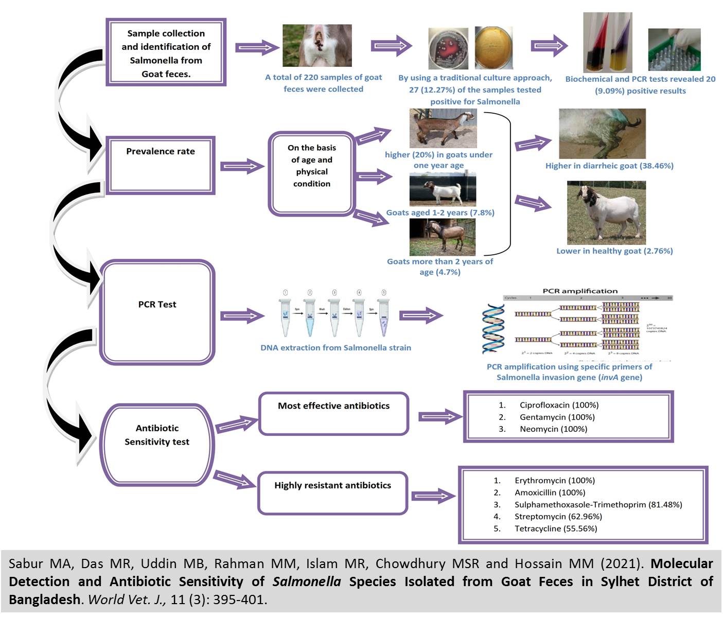
Molecular Detection and Antibiotic Sensitivity of Salmonella Species Isolated from Goat Feces in Sylhet District of Bangladesh
Sabur MA, Das MR, Uddin MB, Rahman MM, Islam MR, Chowdhury MSR and Hossain MM.
World Vet. J. 11(3): 395-401, 2021; pii:S232245682100051-11
DOI: https://dx.doi.org/10.54203/scil.2021.wvj51
ABSTRACT: The present study aimed at the molecular detection of Salmonella species from feces of goats and the characterization of the isolated Salmonella by biochemical and antimicrobial sensitivity techniques. A total of 220 goat feces samples were collected, of which 27 (12.27%) were positive for Salmonella by conventional culture methods and 20 (9.09%) by biochemical and PCR techniques. The prevalence was higher in goats under one year of age (20%), compared to older animals aged one to two years (7.8%) and more than two years of age (4.7%), respectively. Moreover, the prevalence of diarrheic goats was significantly higher (38.46%) than healthy animals (2.76%). DNA was extracted from Salmonella strains and amplified by PCR using the specific primers of Salmonella invasion gene (invA gene). The antibiotic sensitivity test indicated that Ciprofloxacin (100 percent sensitivity), Gentamycin (100 percent sensitivity), and Neomycin (100 percent sensitivity) were the most effective antibiotics for the majority of Salmonella isolates. On the other hand, Salmonella isolates were found to have substantially high resistance to Erythromycin (100%), Amoxicillin (100%), Trimethoprim-Sulfamethoxazole (81.48%), Streptomycin (62.96%), and Tetracycline (55.56 percent). Since the rate of Salmonella carriers was relatively high, eating goat meat could increase the risk of foodborne salmonellosis.
Keywords: Antibiotic sensitivity, Goat isolation, PCR detection, Salmonella
[Full text-PDF] [XML] [Google Scholar] [Crossref Metadata] [Scopus ID: 85115401667] [Export from ePrint] [How to Cite]
 Research Paper
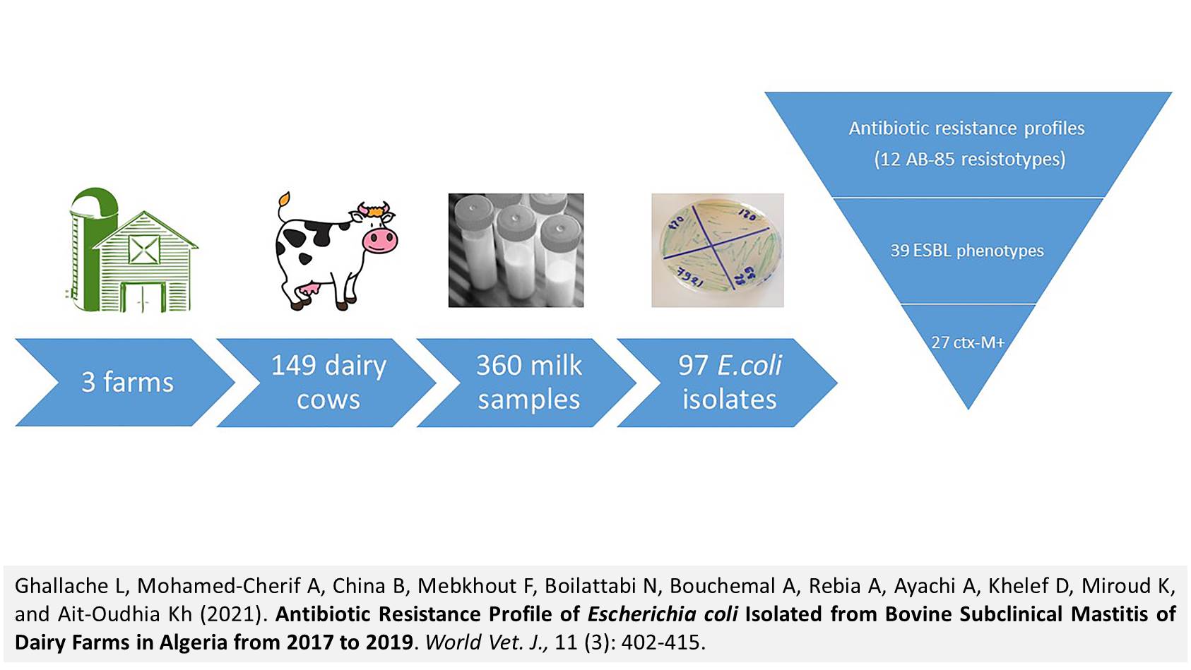
Antibiotic Resistance Profile of Escherichia coli Isolated from Bovine Subclinical Mastitis of Dairy Farms in Algeria from 2017 to 2019
Ghallache L, Mohamed-Cherif A, China B, Mebkhout F, Boilattabi N, Bouchemal A, Rebia A, Ayachi A, Khelef D, Miroud K, and Ait-Oudhia Kh.
World Vet. J. 11(3): 402-415, 2021; pii:S232245682100052-11
DOI: https://dx.doi.org/10.54203/scil.2021.wvj52
ABSTRACT: Mastitis in cows is a major problem in dairy farms leading to a decrease in the quantity and quality of milk. The aim of the present study was to examine the association between the presence of Escherichia coli (E. coli) in milk and the subclinical mastitis, and to characterize the antibiotic resistance profiles of the isolated E. coli. In the current study, a total of 360 cow raw milk samples from three dairy farms of the region of Algiers were analyzed. The analysis period lasted from Spring 2017 to Winter 2019. The California Mastitis Test (CMT) was applied to detect subclinical mastitis. The E. coli strains were isolated from milk using conventional bacteriological methods. The antibiotic resistance profile of the isolated E. coli strains to 12 different antibiotics was tested using the disk diffusion method. On β-lactamase-producing strains, a double diffusion test was applied to identify the Extended-spectrum β-lactamase (ESBL) phenotype. Finally, the ctXx-M genes were amplified by PCR. Two-thirds (66.4%) of the milk samples were positive for the CMT test. A total of 97 E. coli strains were isolated from the milk samples, their resistance to antibiotics was tested, and 3.1% of the strains were resistant to trimethoprim-sulfamethoxazole, 6.2% to chloramphenicol, 12.3% to gentamicin, 13.4% to colistin, 23.3% to amoxicillin/clavulanate, 31.9% to kanamycin, 39.2% to enrofloxacin, 51.5% to cefotaxime, 52% to tetracycline, 57.7% to ampicillin, 74.3% to nalidixic acid, and 75.3% to amoxicillin. Furthermore, most of the E. coli strains (92.8%) were resistant to more than one antibiotic with a Multiple Antibiotic Resistance index ranging from 0 to 0.8. The 50 strains resistant to cefotaxime were analyzed for an ESBL phenotype. 39 of them (78%) were positive to the double-disk synergy test. Among the 39 ESBL positive strains, 27 (69.2%) were confirmed for the presence of a CTX-M gene by PCR. The present study showed that multiple drug-resistant E. coli, including ESBL-carriers, were frequently isolated from the milk of dairy cows in Algeria. The results underlined that the use of antibiotics on farms must be reasoned to avoid the spread of resistant strains in animals and human populations.
Keywords: Antibiotic Resistance, Cows, CTX-M gene, Escherichia coli, Milk, Subclinical Mastitis
[Full text-PDF] [XML] [Google Scholar] [Crossref Metadata] [Scopus ID: 85115368187] [Export from ePrint] [How to Cite]
 Research Paper
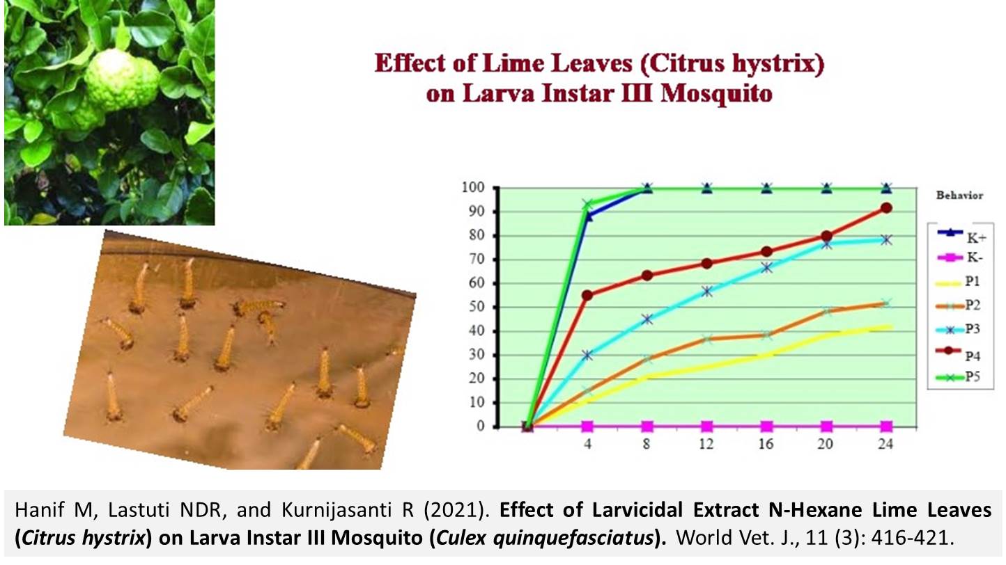
Effect of Larvicidal Extract N-Hexane Lime Leaves (Citrus hystrix) on Larva Instar III Mosquito (Culex quinquefasciatus)
Hanif M, Lastuti NDR, and Kurnijasanti R.
World Vet. J. 11(3): 416-421, 2021; pii:S232245682100053-11
DOI: https://dx.doi.org/10.54203/scil.2021.wvj53
ABSTRACT: Culex quinquefasciatus mosquitoes are a vector of transmission of several types of diseases, such as filariasis, Japanese encephalitis, and dirofilariasis. Larval control is the key strategy of disease control programs caused by vectors around the world because it can prevent larvae to enter the adult stage. Control of mosquito larvae that are often used is chemically controlled. Citrus hystrix is a natural plant and contains chemical compounds that have biological activity, such as flavonoids, carotenoids, and limonoids. The present study aimed to determine the effect of citrus leaf extract (Citrus hystrix) on the mortality of the larva Culex quinquefasciatus. The method used in the current study was a laboratory experimental study method with the experimental design using a completely randomized design. The research plot carried out was the rearing larvae of Culex quinquefasciatus, the manufacture of citrus leaf extract, the manufacture of larvicidal material, and the observation phase after treatment. Extract N-hexane omplet name hystrix leaf has high toxicity because it contains essential oils, flavonoids, alkaloids, terpenoids, saponins, and Limonoids. The Optimal concentration of N-hexane extracts of citrus leaves that lead to larval mortality of Culex quinquefasciatus in vitro amounted to 93.33% on 4 hours of observation with a concentration of 4000 ppm. Extract N-hexane lime leaf has a larvicidal effect on the larva Culex quinquefasciatus in vitro.
Keywords: Citrus hystrix, Culex quiquefasciatus, Extract, Mosquitoes, Larvicide
[Full text-PDF] [XML] [Google Scholar] [Crossref Metadata] [Scopus ID: 85120079025] [Export from ePrint] [How to Cite]
 Research Paper
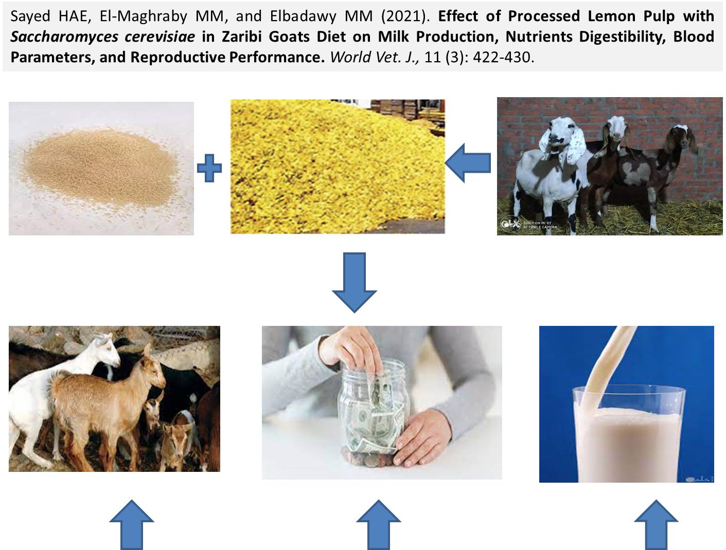
Effect of Processed Lemon Pulp with Saccharomyces cerevisiae in Zaribi Goats Diet on Milk Production, Nutrients Digestibility, Blood Parameters, and Reproductive Performance
Sayed HAE, El-Maghraby MM, and Elbadawy MM.
World Vet. J. 11(3): 422-430, 2021; pii:S232245682100054-11
DOI: https://dx.doi.org/10.54203/scil.2021.wvj54
ABSTRACT: The aim of the present study was to evaluate the effect of replacing concentrate feed (CFM) mixture with different levels of treated lemon pulp (TLP) on milk production and composition, nutrients digestibility, and blood parameters of Zaribi goats. A total of 24 Zaribi female goats at the end of the pregnant period were divided into three groups of eight according to live weight. Concentrate feed mixture and clover hay at a rate of 50:50 were offered twice a day. Treatedlemon pulp replaced CFM at rates 0%, 25%, and 50 % in diets of R1, R2, and R3 treatment groups, respectively. The feeding trial lasted 90 days. Dry matter digestibility and Nitrogen free extract digestibility were significantly raised by the increased level of TLP in groups R2 (72.37% and 70.36%) and R3 (72.28% and 70.30%), compared to (70.99% and 68.51%) in R1, respectively. The same trend was observed for organic matter digestibility (OMD), crude protein digestibility (CPD), crude fiber digestibility (CFD), and ether extract digestibility (EED) with R3, compared with either R1 or R2. However, there were insignificant differences between R1 and R2 in terms of OMD, CPD, and EED but CFD was significantly higher in R2, compared to R1. There was a significant increase in the milk yield by the increased level of TLP in the diet and for R2 (1448.4g/h/d) and R3(1558.7g/h/d), while it was reported as 1377.6 g/h/d in the control group. Feeding dams on the R3 diet had a significant effect on improving total antioxidant capacity by 63.5%, compared with control. The results of the present study indicated that the replacement of CFM by TLP decreased the costs of feed and increase milk production for the replacement level by 50% (R3). Moreover, no adverse effects were noticed on nutrients digestibility and blood parameters of the investigated samples.
Keywords: Blood parameters, Lemon pulp, Milk production, Saccharomyces cerevisiae
[Full text-PDF] [XML] [Semantic Scholar] [Crossref Metadata] [Scopus ID: 85120061377] [Export from ePrint] [How to Cite]
 Short Communication
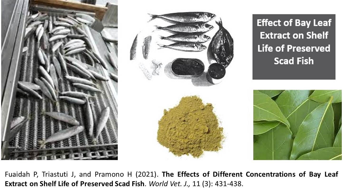
The Effects of Different Concentrations of Bay Leaf Extract on Shelf Life of Preserved Scad Fish
Fuaidah P, Triastuti J, and Pramono H.
World Vet. J. 11(3): 431-438, 2021; pii:S232245682100055-11
DOI: https://dx.doi.org/10.54203/scil.2021.wvj55
ABSTRACT: Scad fish (Decapterus kurroides) is the most productive fish species. To provide the added value, fishermen can process scad fish into various forms of processed products, such as preserved fish, which extend the shelf life. In order to extend the shelf life of canned fish, natural preservatives could be used that contain antimicrobial agents, such as bay leaves. The present study aimed to determine the effects of bay leaf extract in different concentrations on the shelf life of preserved scad fish. In the present study, the completely randomized design was used as an experimental research method. The treatments given differed in terms of the concentration of the bay leaf extract. The groups were treated as the scad fish without bay leaf extract (A), scad fish soaked in 6% of bay leaf extract (B), scad fish soaked in 7% of bay leaf extract (C), scad fish soaked in 8% of bay leaf extract (D), and scad fish soaked in 9% of bay leaf extract (E). The analyzed parameters included the total plate count, pH, and water level test using Analysis of Variance (ANOVA). The supportive parameters observed included an organoleptic test. The results indicated that the lowest total bacterial count from the beginning to the end of the experiment (18 hours) occurred on treatment C (7% of bay leaf extract addition), which was 1.54 × 10³ to 5.85 × 10⁶. Regarding the water level test from the beginning to the end of the experiment, treatment A (control) was not significantly different from other treatments. The difference in the concentration of bay leaf extract on scad fish effectively inhibited the growth of bacterial colonies. In conclusion, adding bay leaf extract to scad fish could inhibit bacteria for up to 12 hours. Treatment C (7% of bay leaf extract) gave the best results since this concentration level could inhibit the bacteria on scad fish.
Keywords: Bay leaf, Preservation, Scad fish, Shelf life
[Full text-PDF] [XML] [Google Scholar] [Crossref Metadata] [Scopus ID: 85120048844] [Export from ePrint] [How to Cite]
 Research Paper
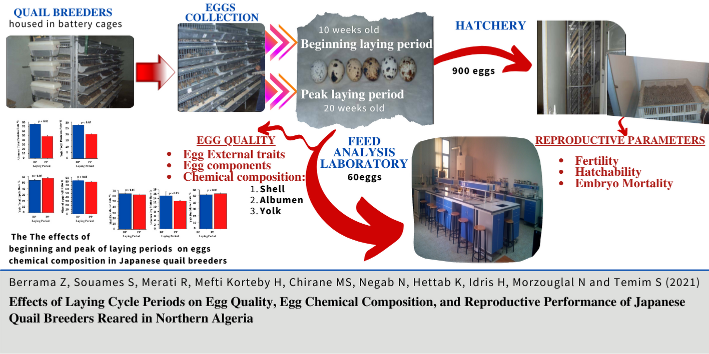
Effects of Laying Cycle Periods on Egg Quality, Egg Chemical Composition, and Reproductive Performance of Japanese Quail Breeders Reared in Northern Algeria
Berrama Z, SouamesS, Merati R, Korteby HM, Chirane MS, Negab N, Hettab K, Idris H, Morzouglal N, and Temim S.
World Vet. J. 11(3): 439-447, 2021; pii:S232245682100056-11
DOI: https://dx.doi.org/10.54203/scil.2021.wvj56
ABSTRACT: Egg quality traits in quail breeders depend on various factors which may influence embryo survival during incubation, affecting the chicks' production and quality. The current study aimed to determine the effect of the laying period on the external and internal quality of the egg, the chemical composition of albumen, yolk, and eggshell as well as the reproductive parameters of quail breeders. A total of 450 quails (Coturnix japonica) obtained from the same hatchery, were reared in a battery cage with a sex ratio of 1 male to 3 females. All the quails were subjected to standard breeding conditions and fed a balanced laying diet. A total of 960 eggs were collected at two different periods of the laying cycle, the beginning period of the laying (BP indicating 10 weeks of quail age) and the peak period of laying (PP showing 20 weeks of quail age). At each laying period, 30 eggs were used to analyze the various parameters of egg quality, and 450 eggs were randomly selected to assess the reproductive performances. Overall, no differences in the external quality of eggs, such as egg length, egg width, and the egg shape index, were recorded between the two laying periods. Apart from albumen weight that tended to be higher at the peak laying period, the shell and the yolk weights were not affected by the period of laying. Additionally, Japanese quail tend to deposit similar proportions of shell, albumen, and yolk at the two periods of laying. Likewise, the rate of dry matter of the three egg components, shell mineral concentrations, and yolk fat concentrations did not show any noticeable variation with the laying period. The most significant effect of the laying period was related to the potential reduction in the total protein content of the albumen and the yolk of eggs laid at the peak period of laying. Finally, the laying period did not significantly affect the fertility and hatchability rate of the incubated quail eggs but slightly improved the embryonic mortality rate during the peak laying phase.
Keywords: Albumen, Coturnix japonica, Eggshell, Fertility, Quail
[Full text-PDF] [XML] [Google Scholar] [Crossref Metadata] [Scopus ID: 85120049011] [Export from ePrint] [How to Cite]
 Research Paper
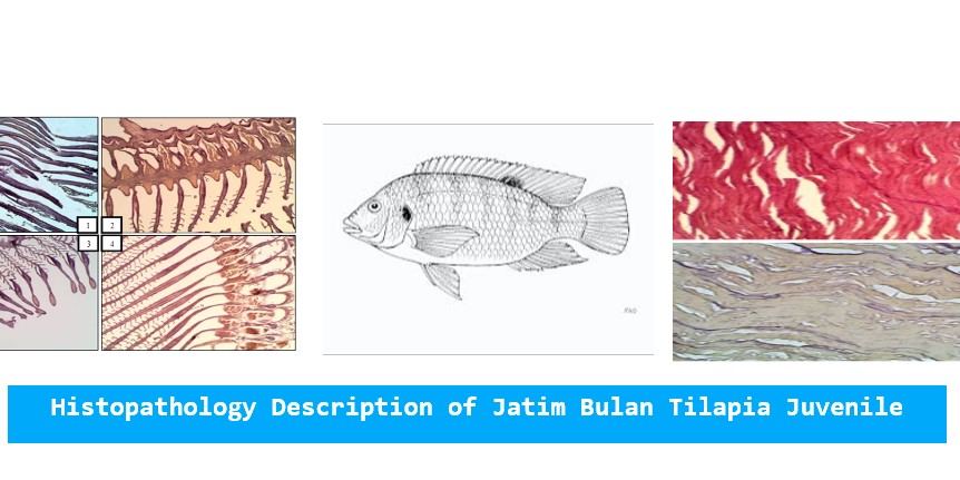
The Role of Salinity in Histopathology Description of Jatim Bulan Tilapia Juvenile (Oreochromis niloticus) Exposed by Lead (PB)
Batista FR, Triastuti J, and Pursetyo KT.
World Vet. J. 11(3): 448-455, 2021; pii:S232245682100057-11
DOI: https://dx.doi.org/10.54203/scil.2021.wvj57
ABSTRACT: The decrease in the number of lands for aquaculture will cause the freshwater fish aquaculture, especially tilapia is reared. As one of the efforts to survive, the breeders use the coastal area to anticipate, so the freshwater fish can adapt to the sea waters. Heavy metal pollution near the coasts (or in the coastal waters) has great potential impacts on the environment. The present study aimed to find out the effect of Lead (Pb, heavy-metal) exposure on the Jatimbulan Juvenile tilapia with the different salinity towards histopathology description of the gill, intestine, and the skin tissues. The method used was an experimental method with a completely randomized design using six treatments and three times repetition. The observed parameters included the changes in histopathology description of gill, intestine, and skin organs of Jatimbulan juvenile tilapia. Kruskal-Wallis scoring method was used for data analysis and was continued by Mann-Whitney. The result of the current study showed the effect of salinity towards histopathology description of gill, intestine, and skin of Jatimbulan juvenile tilapia. The effect of changes was proven with the histopathology description in the form of the damage of edema, hyperplasia, and necrosis on the gill tissue; the damage of edema, atrophy, and necrosis on the gill tissue as well as the damage of edema, atrophy, hemorrhagic and necrosis on the gill tissue. From the result of the current study which has been conducted, it can be concluded that the heavy-metal exposure by lead was 0.03 ppm on the salinity of 10 ppt and 20 ppt. given the significant effect on the histopathology description of gill, intestine, and skin of Jatimbulan juvenile tilapia.
Keywords: Histopathology, Lead, Metal, Oreochromis niloticus, Salinity
[Full text-PDF] [XML] [Google Scholar] [Crossref Metadata] [Scopus ID: 85120042019] [Export from ePrint] [How to Cite]
 Research Paper
In Vitro Antibiotic Activity of red Shallot (Allium ascalonicum), Mulberry (Morus indica), and Marigold (Tagetes erecta) Extracts against Streptococcus pyogenes
Mekvimol T, Chaipunna Ch, Poonthong G, and Pumipuntu N.
World Vet. J. 11(3): 456-461, 2021; pii:S232245682100058-11
DOI: https://dx.doi.org/10.54203/scil.2021.wvj58
ABSTRACT: Bacterial infection is a major global health concern. One of the critical problems is the widespread of antimicrobial-resistant bacteria from inappropriate and prolonged use of antimicrobial agents in both humans and animals. Plant extracts might afford the chance to replace antibiotic drugs and reduce the emerging of antimicrobial-resistant bacteria. This study aimed to examine the antibiotic activity of ethanolic crude extracts of some Thai medicinal plants grouping in their parts as whole onions of red shallot (Allium ascalonicum), petals of marigold (Tagetes erecta), mulberry leaves, and root barks (Morus indica) to inhibit the growth of Streptococcus pyogenes. The antibiotic activities of the crude extract of three Thai medicinal plants using absolute ethanol were trialed against Streptococcus pyogenes using the disk diffusion method. Erythromycin and Ceftriaxone discs were chosen to be positive control standards as the representative of antibiotic drugs. Each dried plant extracts was prepared to test the inhibition with a concentration at 25, 50, and 75 mg/mL stock solution. The results showed that three groups from all testing groups of Thai medicinal plant extracts had the potential of antibiotic activity against S. pyogenes. The highest antibiotic activity against S. pyogenes was detected from whole onion extract red shallot followed by the extract of the mulberry leaves and root barks of mulberry strain Nakhon Ratchasima 60 (Nak 60) while the extract of marigold petal did not present antibiotic activity. The results revealed that crude extract of those two Thai medicinal plants, including red shallot and mulberry, had antibiotic activity against bacterial growth of S. pyogenes in the experiment and these medical Thai plants had potential benefits for developing as alternative treatment agents for S. pyogenes infections in both humans and animals in the future.
Keywords: Antibiotic activity, Ethanolic crude extract, Thai medicinal plants, Streptococcus pyogenes
[Full text-PDF] [XML] [Semantic Scholar] [Crossref Metadata] [Scopus ID: 85120064611] [Export from ePrint] [How to Cite]
 Research Paper
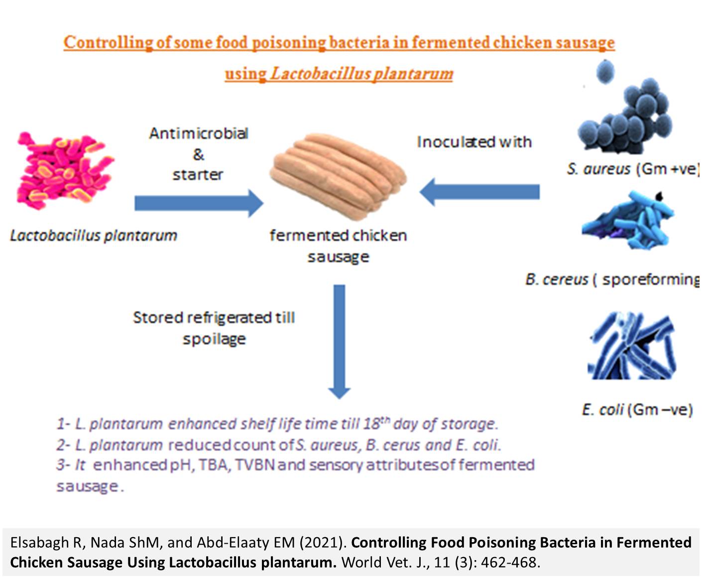
Controlling Food Poisoning Bacteria in Fermented Chicken Sausage Using Lactobacillus plantarum
Elsabagh R, Nada ShM, and Abd-Elaaty EM.
World Vet. J. 11(3): 462-468, 2021; pii:S232245682100059-11
DOI: https://dx.doi.org/10.54203/scil.2021.wvj59
ABSTRACT: Lactobacillus plantarum (L. plantarum) bacteria is generally recognized as safe and widely used in the food industry. The current study aimed to study the antimicrobial effects of L. plantarum against some food poisoning microorganisms, such as Staphylococcus aureus (S. aureus), Bacillus cereus (B. cereus), and Escherichia coli (E. coli) in oriental fermented chicken sausage for 18 days of storage at 4ᵒC. The L. plantarum has broad-spectrum antimicrobial effects that enhance the quality and safety of food products. L. plantarum reduced the count of S. aureus, B. cerus, and E. coli to 1.54, 4.26, and 3.03 Log10, respectively, after 18 days of refrigerated storage. Moreover, there were significant effects of L. plantarum on pH, thiobarbituric acid, total volatile basic nitrogen, and sensory attributes of fermented sausage samples during storage time. It was revealed that L. plantarum enhanced the physic-chemical, sensory attributes, and shelf life of fermented chicken sausage. Moreover, L. plantarum inhibited the inoculated food poisoning bacteria in fermented chicken sausage. In conclusion, it is recommended to use L. plantarum in fermented meat products as a starter and a bio-preservative to enhance the quality of the fermented chicken sausage.
Keywords: Chicken sausage, Food safety, Lactobacillus plantarum, Probiotic
[Full text-PDF] [XML] [Semantic Scholar] [Crossref Metadata] [Scopus ID: 85119897033] [Export from ePrint] [How to Cite]
 Short Communication
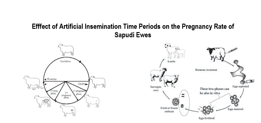
The Effect of the Different Artificial Insemination Time Periods on the Pregnancy Rate of Sapudi Ewes
Putri CD, Ismudiono, and Poetranto ED.
World Vet. J. 11(3): 469-473, 2021; pii:S232245682100060-11
DOI: https://dx.doi.org/10.54203/scil.2021.wvj60
ABSTRACT: Brucellosis Artificial insemination is required to increase the reproduction rate in ruminant breeding. The artificial insemination success rate in sheep only reaches 47.6%, whereas the proposed ideal rate is 70%. One of the factors influencing the artificial insemination success rates in sheep is improper estrus detection, resulting in no fertilization. The present study aimed to determine the effect of different artificial insemination time periods on the pregnancy rates of Sapudi ewes. The research design was based on a completely randomized design. A total of 20 female Sapudi sheep were divided into four treatment groups with five repetitions (for each group). In addition, the observed variables were artificial insemination time in Sapudi ewes. Estrus synchronization in ewes was conducted by injecting PGF2α. The results of the research indicated that ewes subjected to artificial insemination 6, 12, 18, and 24 hours after estrus had a pregnancy rate of 20%, 100%, 60%, and, 60%, respectively. It can be concluded that the time differences in artificial insemination significantly influence the pregnancy rate in Sapudi ewes’.
Keywords: Artificial insemination, Estrus, Pregnancy rate, Sapudi ewes
[Full text-PDF] [XML] [Google Scholar] [Crossref Metadata] [Scopus ID: 85120065728] [Export from ePrint] [How to Cite]
 Research Paper
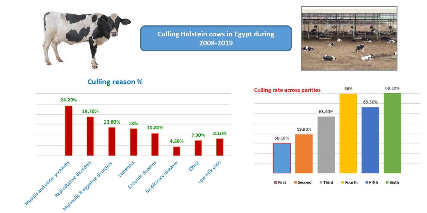
Milk Production and Reproductive Performance of Retained and Culled Cows in a Large Holstein Herd in Egypt
Fahim NH, Ibrahim MA-AM, Amin AH, and Sadek RR.
World Vet. J. 11(3): 474-483, 2021; pii:S232245682100061-11
DOI: https://dx.doi.org/10.54203/scil.2021.wvj61
ABSTRACT: The study aimed to identify the culling reasons of Holstein cows raised in a large commercial herd in Egypt with emphasis on the performance of retained and culled cows. A total of 31534 complete lactation records for 10994 cows calved from 2008 to 2019 were used. The overall rate of culling per lactation was 61.1%. Involuntary culling represented 92% of all culling cases. The reasons for culling included mastitis and udder problems (24.2%), reproductive disorders (18.7%), metabolic and digestive disorders (13.6%), lameness (13%), endemic diseases (10.8%), low milk yield (8.1%), respiratory diseases (4.3%) and unknown causes (7.3%). Means of 305-day milk yield and daily milk yield were significantly lower in culled cows than the retained ones. On the other hand, no significant differences were observed between culled and retained cows for days open and the number of services/conception. The high involuntary culling rate of Holstein under the Egyptian conditions revealed that management practices regarding mastitis prevention and reproductive efficiency should be improved.
Keywords: Culling reasons, Egypt, Holstein, Milk production, Reproductive performance
[Full text-PDF] [XML] [Google Scholar] [Crossref Metadata] [Scopus ID: 85120069604] [Export from ePrint] [How to Cite]
 Short Communication
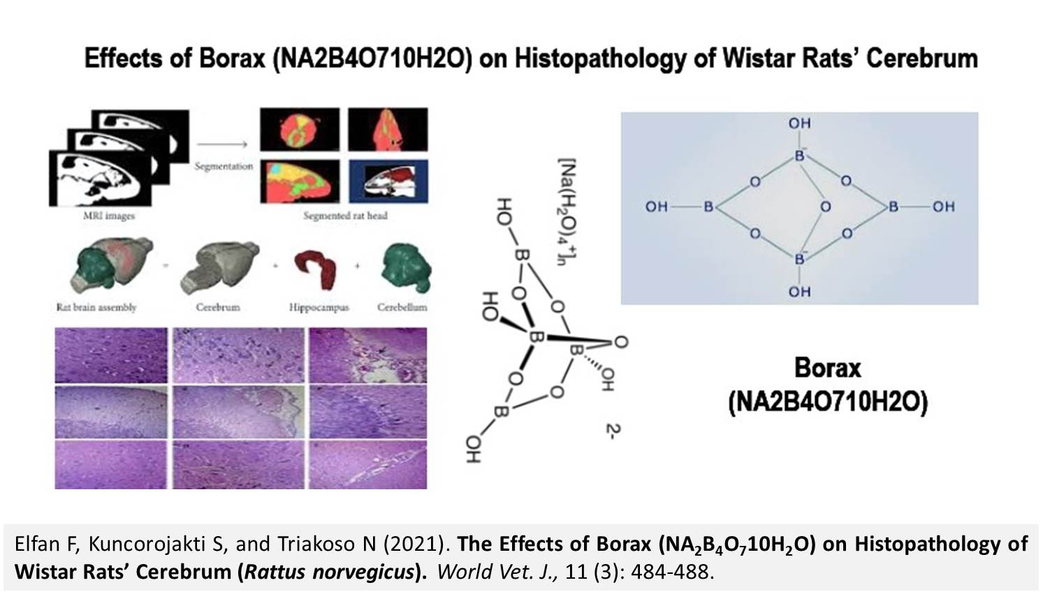
The Effects of Borax (NA2B4O710H2O) on Histopathology of Wistar Rats’ Cerebrum (Rattus norvegicus)
Elfan F, Kuncorojakti S, and Triakoso N.
World Vet. J. 11(3): 484-488, 2021; pii:S232245682100062-11
DOI: https://dx.doi.org/10.54203/scil.2021.wvj62
ABSTRACT: The present study aimed to determine the effects of borax (Na2B4O7.10H2O) addition on the changes of histological cerebrum imaging in the brains of white mice (Rattus norvegicus). The current research was an experimental study with randomization of 24 white mice that were divided into four treatment groups with five replications. Borax was dissolved for each treatment with a dose of 19 mg/mouse/day, 26 mg/mouse/day, and 37 mg/mouse/day, and it was administered orally for 14 days. Then, it was analyzed statistically using the Kruskal-Wallis test. The statistical analysis results suggested that there were significantly different results in each treatment group. The control treatment with an administration dose of 26 mg/rat/day had a significantly different result in the worst cloudy swelling degeneration of cerebrum in histopathology imaging on Wistar rats (Rattus norvegicus). Using the Mann-Whitney test, it was found that the dose of borax at 37 mg/rat/day led to significant difference, compared to the other treatment groups, which means that 37 mg/rat/day of borax caused the worst pyramidal cell necrosis in histopathology imaging of the cerebrum on white mice. Borax exposure on Wistar rats (Rattus norvegicus) can cause cloudy swelling at a dose of 26mg/head/day, and pyramidal cell necrosis at a dose of 37 mg/head/day.
Keywords: Borax, Cerebrum, Cloudy swelling, Necrosis
[Full text-PDF] [XML] [Google Scholar] [Crossref Metadata] [Scopus ID: 85120046694] [Export from ePrint] [How to Cite]
 Research Paper
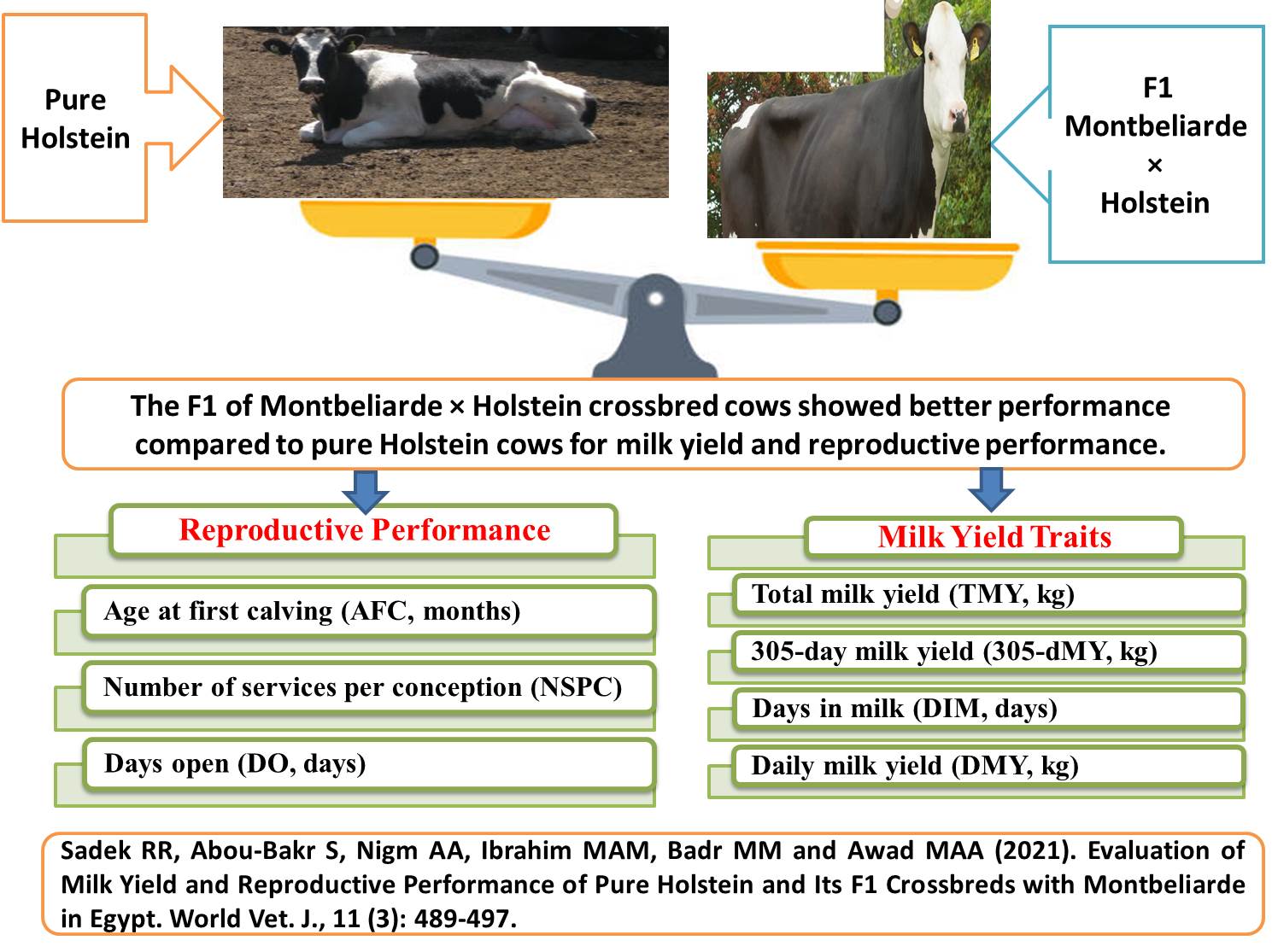
Evaluation of Milk Yield and Reproductive Performance of Pure Holstein and Its F1 Crossbreds with Montbeliarde in Egypt
Sadek RR, Abou-Bakr S, Nigm AA, Ibrahim MAM, Badr MM and Awad MAA.
World Vet. J. 11(3): 489-497, 2021; pii:S232245682100063-11
DOI: https://dx.doi.org/10.54203/scil.2021.wvj63
ABSTRACT: The present study was carried out to compare the milk yield and reproductive performance of pure Holstein (HO) cows with those of their first generation (F1) crossbreds with Montbeliarde cows (MO) in four commercial dairy herds under Egyptian conditions. Data used in the current study comprised 2268 records for the first four lactations of 531 HO and 536 MO × HO F1 crossbred cows during the period between 2012 and 2020. Data were analyzed using the least squares method by XLSTAT software. The MO × HO crossbred cows were significantly superior compared with pure HO cows for 305-day milk yield, scoring 9210 ± 96 kg versus 7987 ± 149 kg. Moreover, MO × HO F1 crossbred cows had a significantly higher daily milk yield (30.0 ± 0.45 kg) than pure HO cows (25.9 ± 0.52 kg). However, pure HO cows had significantly greater days in milk (399 ± 6 days) than MO × HO crossbred cows (341 ± 5.2 days). With regard to reproductive performance, MO × HO F1 crossbred cows had significantly less number of services per conception and days open than pure HO cows (2.6 ± 0.16 vs. 3.7 ± 0.18) and (132 ± 5.2 days vs. 190 ± 6 days), respectively. However, the statistical difference between MO × HO F1 crossbred cows and pure HO cows for age at first calving was not significant (22.9 ± 0.11 vs. 23.1 ± 0.15 months, respectively). It can be concluded that under Egyptian subtropical conditions, the first generation of MO × HO crossbred cows exhibit better performance, compared to pure HO cows in milk yield and reproductive traits. These findings could provide an effective strategic option for the genetic improvement of dairy cattle in hot subtropical regions.
Keywords: Crossbreeding, Egypt, Holstein, Milk Yield, Montbeliarde, Reproduction
[Full text-PDF] [XML] [Google Scholar] [Crossref Metadata] [Scopus ID: 85120070427] [Export from ePrint] [How to Cite]
 Short Communication
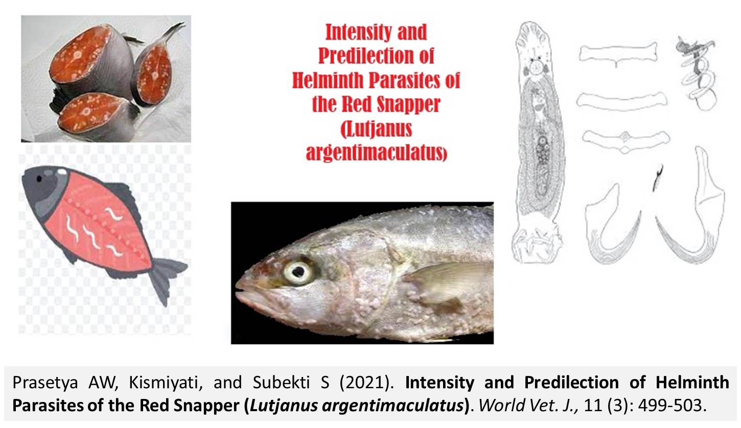
Intensity and Predilection of Helminth Parasites of the Red Snapper (Lutjanus argentimaculatus)
Prasetya AW, Kismiyati, and Subekti S.
World Vet. J. 11(3): 498-503, 2021; pii:S232245682100064-11
DOI: https://dx.doi.org/10.54203/scil.2021.wvj64
ABSTRACT: Marine fish, particularly the red snappers, are often exposed to helminth parasitic infestation. As a result of the parasitic infestation, the fish population, the fish weight, and the morphological changes in the fish are shrinking. The present research aimed to find out the intensity and predilection of the helminth ectoparasites over the infection of the red snapper (Lutjanus argentimaculatus) and employed the survey method for this purpose. The sampling was carried out by the purposive sampling technique. The sample obtained consisted of 30 fish, 20% of the total red snapper population of 150 fish reared in the floating net cages of Balai Besar Perikanan Budidaya Laut Lampung. The intensity of the fish infestation by a mixture of Haliotrema epinepheli and Benedenia epinepheli was 132.5 individuals/fish. Neobenedenia girellae and Haliotrema epinepheli infected fish with an intensity of 149.41 individuals/fish. The 66.7% of Benedenia epinepheli had a predilection for the dorsal fin, and 33.3% for the anal fin. In Neobenedenia girellae, 57.1% had a predilection for the body surface, 37.2% for the head surface, and 5.7% for the dorsal fin. In conclusion, all sampled fish were positively infected with helminth ectoparasites, including Neobenedenia girellae, Haliotrema epinepheli, and Benedenia epinepheli.
Keywords: Ectoparasite, Helminths, Infestation, Red snapper
[Full text-PDF] [XML] [Google Scholar] [Crossref Metadata] [Scopus ID: 85120074705] [Export from ePrint] [How to Cite]
 Research Paper
Cross Reaction of Haemonchus contortus Protein with Toxocara vitulorum Anti-L2 Serum Using Western Blot Technique
Asmorowati RW, Kusnoto, and Eliyani H.
World Vet. J. 11(2): 504-509, 2021; pii:S232245682100065-11
DOI: https://dx.doi.org/10.54203/scil.2021.wvj65
ABSTRACT: In the adult stage, Haemonchus contortus worms infect the abomasum host causing anemia and even death in animals. However, identifying the H. contortus protein can be used as a reference for the diagnosis of diseases. The diagnosis is performed by serological cross-reaction between H. contortus protein and anti-L2 Toxocara vitulorum (T. vitulorum) serum using the western blot technique. The main purpose of the current research was to identify the cross-reaction between H. contortus proteins and anti-L2 T. vitulorum serum using the western blot technique. T. vitulorum worms were collected from the intestine of cattle and H. contortus worms were collected from the abomasum of goats. The first step was making antibodies by oral infection of rats with infective eggs (L2) of T. vitulorum. The blood was taken 21 days after infection. Then, the blood was centrifuged at 1500 rpm for 10 minutes to get the serum. The second step was making homogenates from the whole worm extract of H. contortus. After crushing the worms, it was centrifuged at 5000 rpm for 15 minutes and the supernatant was taken. The supernatant was then analyzed using Sodium Dodecyl Sulphate Polyacrylamide Gel Electrophoresis (SDS-PAGE) with coomassie brilliant blue staining. The third step was the analysis of H. contortus protein with serum anti-L2 T. vitulorum using the western blot technique. From the H. contortus homogenates analysis using SDS-PAGE, 16 protein bands were obtained. The cross-reactions were 141.3, 81.3, 64. 6, 51.3, 46.8, and 38 kDa. The data from cross-reactions suggested that the H. contortus protein cannot be used as a diagnostic material. It is serologically Haemonchosis because it caused false positives with diagnostic Toxocariasis.
Keywords: Cross reaction, Haemonchus contortus, SDS-PAGE, Toxocara vitulorum, Western blot
[Full text-PDF] [XML] [Google Scholar] [Crossref Metadata] [Scopus ID: 85120089399] [Export from ePrint] [How to Cite]
 Review
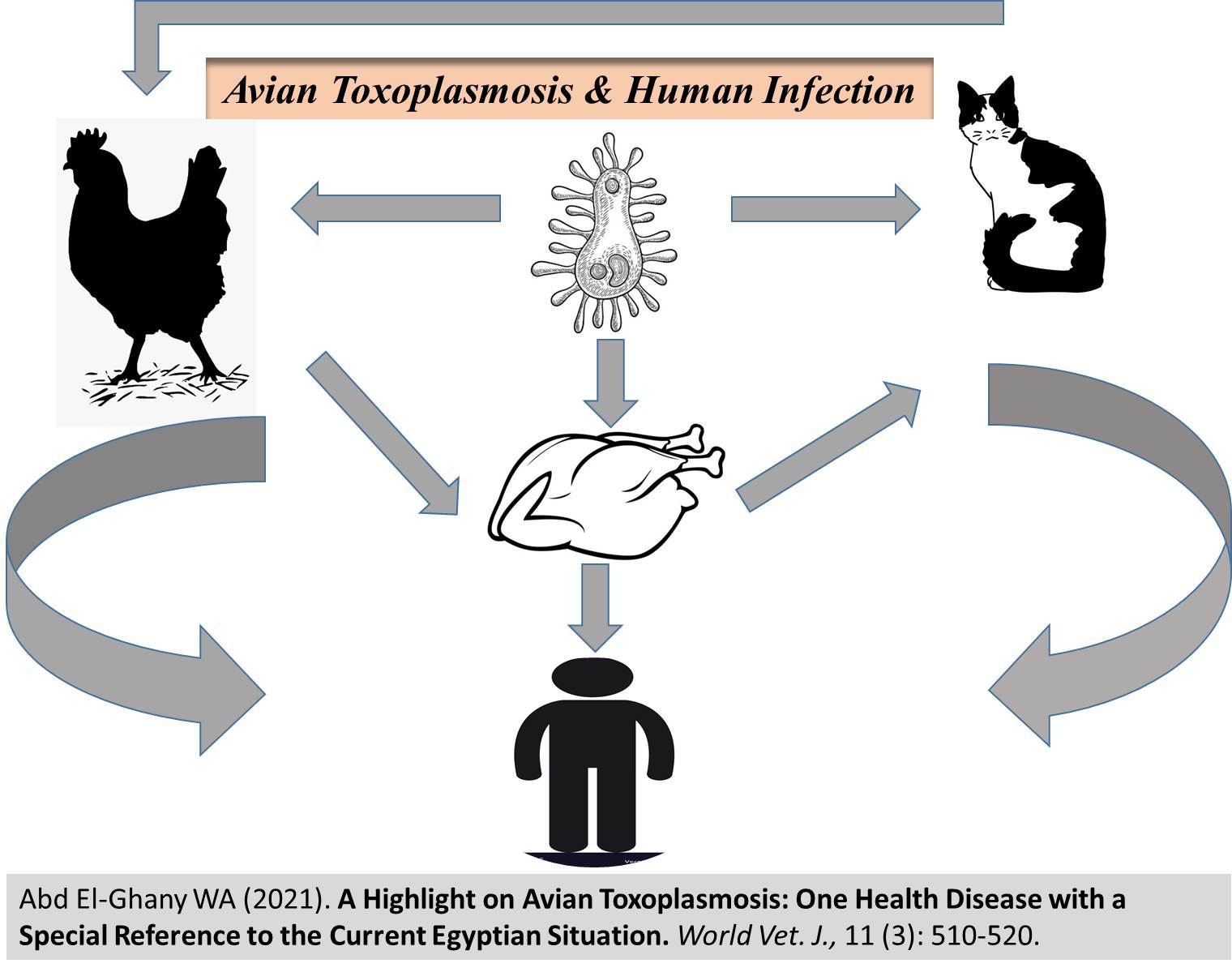
A Highlight on Avian Toxoplasmosis: One Health Disease with a Special Reference to the Current Egyptian Situation
Abd El-Ghany WA.
World Vet. J. 11(2): 510-520, 2021; pii:S232245682100066-11
DOI: https://dx.doi.org/10.54203/scil.2021.wvj66
ABSTRACT: This review article was developed to the infection of avian species with Toxoplasma gondii (T. gondii), diagnosis, pet bird and human infection, and control methods with a special reference to the current status of infection among the Egyptian poultry farms and population. Toxoplasmosis is a zoonotic disease caused by a unicellular, protozoan parasite T. gondii. Different domesticated and wild animals, as well as birds can harbor T. gondii and may be a potential source of infection to humans. Avian species could be infected with T. gondii through the ingestion of contaminated food, soil, and water with oocysts shed in the excreta of infected animals, especially cats. Poor sanitation and hygienic conditions increase the risk of infection. Consumption of food or water, as well as undercooked poultry meat or meat products containing the oocysts of the parasite, are the main sources of human infection with T. gondi. Diagnosis of T. gondii in the infected host depends on the serological detection of specific antibodies and molecular detection of the parasite. Microscopic demonstration of the oocysts and other developmental stages of the parasite in the intestine, liver, brain, and skeletal muscles tissues is another means for rapid diagnosis. Generally, a high prevalence of the disease is also reported in pet birds. Toxoplasmosis in humans is associated with abortion, congenital disorders, stillbirth, and other complications, especially in immunocompromised patients. Application of hygienic measures, as well as public awareness, are essential for the prevention and control of toxoplasmosis. In different Egyptian governorates, a high prevalence of T. gondii has been detected in animals, birds, and humans. High incidence of infection was recorded due to the contact with Toxoplasma oocysts shed mainly from infected cats or other carriers. Egyptian chicken and turkey flocks and backyard birds revealed the presence of different developmental stages of the parasite and even its antibodies. In addition, human populations showed signs of toxoplasmosis with severe complications.
Keywords: Bird, Egypt, Human, Toxoplasma gondii, Zoonosis
[Full text-PDF] [XML] [Semantic Scholar] [Crossref Metadata] [Scopus ID: 85120079422] [Export from ePrint] [How to Cite]
Previous issue | Next issue | Archive
![]() This work is licensed under a Creative Commons Attribution 4.0 International License (CC BY 4.0).
This work is licensed under a Creative Commons Attribution 4.0 International License (CC BY 4.0).