 |
|  |
| 
Previous issue | Next issue | Archive
Volume 7 (1); March 25, 2017 [Booklet]
Research Paper
Prevalence and Associated Risk Factors of Bovine Schistosomiasis in Northwestern Ethiopia.
Kassahun Gebremeskel A, Tayelgn Simeneh S and Addisu Mekuria Sh.
World Vet. J. 7(1): 01-04, 2017; pii:S232245681700001-7
DOI: http://dx.doi.org/10.5455/wvj.20170287
ABSTRACT
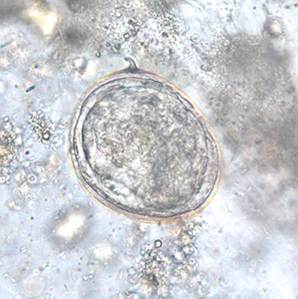
Schistosomiasis is a parasitic disease caused by microorganisms from the genus Schistosoma. It has a huge negative impact on both economy and health worldwide. In this paper a cross-sectional study was conducted to evaluate the prevalence and associated risk factors of bovine schistosomiasis in north western Ethiopia with the objective of providing detailed information on bovine schistosomiasis prevalence in relation to animal and ecological related risk factors. The sampled animals were categorized under four associated risk factors which include: origin, sex, body condition score and age. Fecal samples were randomly collected from a total of 289 animals and Schistosoma’s eggs were identified by sedimentation technique. 69 (23.9%) fecal samples were revealed positive for Schistosoma. The highest (29.8%) prevalence rate was recorded at Keltafa district followed by Lalibella (25.9 %), Korench (19.1%) and kurbiha (14.0%). Animals categorized under medium body condition score has a relative high prevalence (25.7%) followed by poor (24.3%) and good body condition (21.7%) animals. In conclusion, the prevalence recorded among different selected study districts, sex, body condition score and age groups shows some degree of variability and insignificant (p>0.05), which resulted from the difference in abundancy of marshy areas and rivers, animal’s immunity and types of management system. Despite these variability factors, the disease has a great socio-economic impact that needs intervention.
Key words: Bovine, Prevalence, Schistosomiasis, Ethiopia
[Full text-PDF] [XML] [Import into EndNote] [Citations on Google Scholar]
 Research Paper
Resistant Gene of Pseudomonas Aeruginosa in Mastitic Cattle; Biochemical and Immunological Parameters.
Awad Ibrahim N, El-Metwally Farag VM, Abd-El-Moaty AM and Magdy Atwa S.
World Vet. J. 7(1): 05-13, 2017; pii:S232245681700002-7
DOI: http://dx.doi.org/10.5455/wvj.20170288
ABSTRACT
Mastitis is an important infectious disease of cattle and Pseudomonas aeruginosa (P. aeruginosa) bacteria standout amongst those fundamental causative agents. The present study was intended to assess P. aeruginosa activity isolated from mastitic cattle milk samples in a veterinary hospital. It also was assessed some free radicals and immunological parameters in the milk and serum samples. Control samples were taken from apparently healthy cows, negative California Mastitis test. The results cleared that positive P. aeruginosa isolates were 34 of 100 milk samples. In vitro antibiotic sensitivity test indicated that 79.4%, 70.5% and 58.82% of the isolates completely resisted cefotaxime, penicillin and amikacin respectively. Also, the resistance to meropenem was 11.76% and 8.8% for carbapenem resistant isolate which completely resisted other classes of β-lactams. While enrofloxacin and gentamicin sensitivity reached to 76.47% and 73.5% respectively. The technique of the P.C.R was done for detection of MexR gene (in isolates resisted to more than three antibiotics) and Vim gene (in carbapenem resistant isolates). The biochemical results investigated that the nitric oxide and Mmalondialdehyde antioxidant levels were increased significantly while the cholesterol was decreased significantly in both serum and milk samples. Meanwhile, catalase and lysozyme were changed between groups and total protein and globulin had increased significantly in milk samples only. In conclusion, P. aeruginosa isolates including MexR and blaVim genes showed considerable percent of resistance to carbabenem group and other classes of β-lactam. In addition, the estimated biochemical and immunological parameters were affected in that case of mastitis in cattle. The results may encourage studies which are concerned with antioxidants treatment for mastitis in cattle. It may be a key for decreasing body resistance to antibiotics.
Key words: Mastitis, Cattle, Pseudomonas aeruginosa, PCR, Lysozyme
[Full text-PDF] [XML] [Import into EndNote] [Citations on Google Scholar]
 Research Paper
Isolation and Characterization of Bacterial Species from Respiratory Tracts of Cattle Slaughtered in Addis Ababa City, Central Ethiopia.
Kassahun Gebremeskel A, Sisay Tesema T, Awukew Yegoraw A, Tesfaye Birhanu B and Addisu Mekuria Sh.
World Vet. J. 7(1): 14-20, 2017; pii:S232245681700003-7
DOI: http://dx.doi.org/10.5455/wvj.20170289
ABSTRACT
The present study was an attempt to isolate and identify the diverse bacteria localizing pneumonic lungs and the associated tracheas of 50 slaughtered cattle at Addis Ababa Abattoirs enterprise, central Ethiopia, in both aerobic and anaerobic environments. 158 and 135 bacterial isolate was found in aerobic and anaerobic state, respectively using primary and secondary microbiological testes. Gram positive bacteria were the dominant bacteria in both conditions. The frequency of isolation increased from trachea down to the lung in both state indicating the bacterial role in the progress of bovine pneumonia. Most prevalently isolated bacteria from both aerobics and anaerobics conditions were Staphylococcus species, Bacillus species, Mannheimia haemolytica and Pasteurella multocida. Whereas the Streptococcus species, E.coli, Klebsiella pneumoniae, Actinobacillus species, Micrococcus species, Arcanobacterium, species Neisseriaspecies, Acinetobacter species, Corynebacterium species, Bordetella species, Pseudomonas species, and Rhodococcus equi were among the bacteria isolated.
Key words: Aerobic, Anaerobic, Bacteria, Bovine, Pneumonia
[Full text-PDF] [XML] [Import into EndNote] [Citations on Google Scholar]
 Research Paper
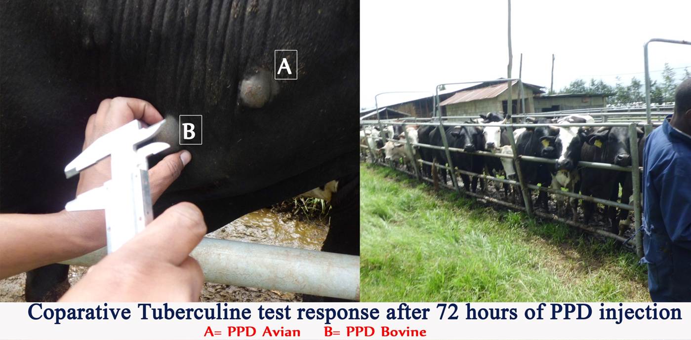
Bovine Tuberculosis Prevalence, Potential Risk Factors and Its Public Health Implication in Selected State Dairy Farms, Central Ethiopia.
Ambaw Endalew M, Deresa B and Ameni Chimdi G.
World Vet. J. 7(1): 21-29, 2017; pii:S232245681700004-7
DOI: http://dx.doi.org/10.5455/wvj.20170290
ABSTRACT
Bovine tuberculosis (BTB), caused by Mycobacterium bovis (M.bovis), is endemic in Ethiopia. However, its magnitude in cattle and human population are not well documented. A cross-sectional study was conducted on 720 apparently healthy dairy cattle kept in three different state owned farms in central Ethiopia to quantify the risk factors and determine the prevalence of BTB using (CIDT) Comparative Intra-Dermal Tuberculin Test from December 2013 to November 2014. Questionnaire survey was used to assess the risk factors and zoonotic implication of BTB. The prevalence of BTB was at 16.53% (95% CI 14.2-18.9) and It was significantly higher in crossbreed (X2= 54.76; P< 0.001; OR=16.1; 95% CI=6.2-41.1) and animals older than 4 years (X2=34.51; P< 0.001, OR =6.22; 95% CI=3.5-11.12). Moreover, the prevalence was also significantly higher in good body conditioned dairy cattle compared to poor body conditioned dairy cattle (X2=29.69; P < 0.001; OR=2.45; 95% CI=1.1-5.7). The prevalence of BTB was also significantly varied among the reproductive status of the dairy cattle (X2 =18.10; P< 0.001).The majority of the respondents consume raw milk (66.1% and raw meat (74.20%) respectively. There was statistically significant variation (X2 =12.51; P< 0.03) in consumption habit between educated and non-educated dairy farm workers. The major risk factors for bovine tuberculosis in this study were breed and age of the dairy cattle. Consumption of raw milk and meat is still a common practice in the study farms. Culling of aged dairy cattle and continuous test and slaughter of infected cattle should be practiced at least in state owned dairy farms to decrease the risk of transmission. In addition to awareness creation of the public particularly the dairy farm workers on the zoonotic nature of tuberculosis is of utmost importance to control bovine tuberculosis.
Key words: Bovine tuberculosis prevalence, Dairy cattle, Farm worker, Risk factor
[Full text-PDF] [XML] [Import into EndNote] [Citations on Google Scholar]
 Research Paper
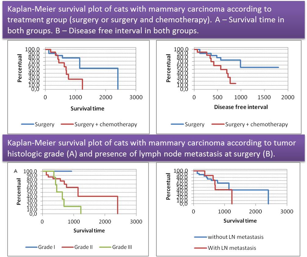
Retrospective Study on Survival Time of Cats with Mammary Carcinomas Undergoing Surgery Alone or with Adjuvant Chemotherapy.
Cunha SCS, Corgozinho KB, Souza HJM, Silva K, Leite J, Mello M and Ferreira AMR.
World Vet. J. 7(1): 30-35, 2017; pii:S232245681700005-7
DOI: http://dx.doi.org/10.5455/wvj.20170291
ABSTRACT
This retrospective study was carried out to evaluate disease free interval (DFI) and survival time of cats with mammary carcinomas that underwent mastectomy (RM) and adjuvant chemotherapy (RMAC) in 35 cats to remove the neoplastic mammary chain and regional inguinal lymphadenectomy. According to performed treatment, the cats were divided into two groups. The RM group (21 cats) received no adjuvant therapy, and the RMAC group (14 cats) received chemotherapy with mitoxantrone or doxorubicin. Histopathological margins were considered complete in all cases. Eight cats had histologically confirmed lymph node involvement at the time of surgery. Three cases were classified as stage I, 21 cases as II and eight cases as III. Nine cats had tumor recurrence (four cats of RM group and five cats of RMAC group) and 12 cats had distant metastasis to the lungs (six cats of each group). Mean and median survival times were 1625 and 2404 days in the RM group, while mean DFI was 815 days. In RMAC group, mean and median survival times were 719 and 690 days, while mean DFI was 549 days. Surgery remains the main treatment and more studies are necessary to evaluate the benefit of adjuvant chemotherapy.
Key words: Feline, Mammary carcinoma, Oncology, Surgery, Chemotherapy
[Full text-PDF] [XML] [Import into EndNote] [Citations on Google Scholar]
Previous issue | Next issue | Archive

This work is licensed under a Creative Commons Attribution-NonCommercial 4.0 International License.


