
Previous issue | Next issue | Archive
![]() Volume 12 (2); June 25, 2022 [Booklet] [EndNote XML for Agris]
Volume 12 (2); June 25, 2022 [Booklet] [EndNote XML for Agris]
 Review
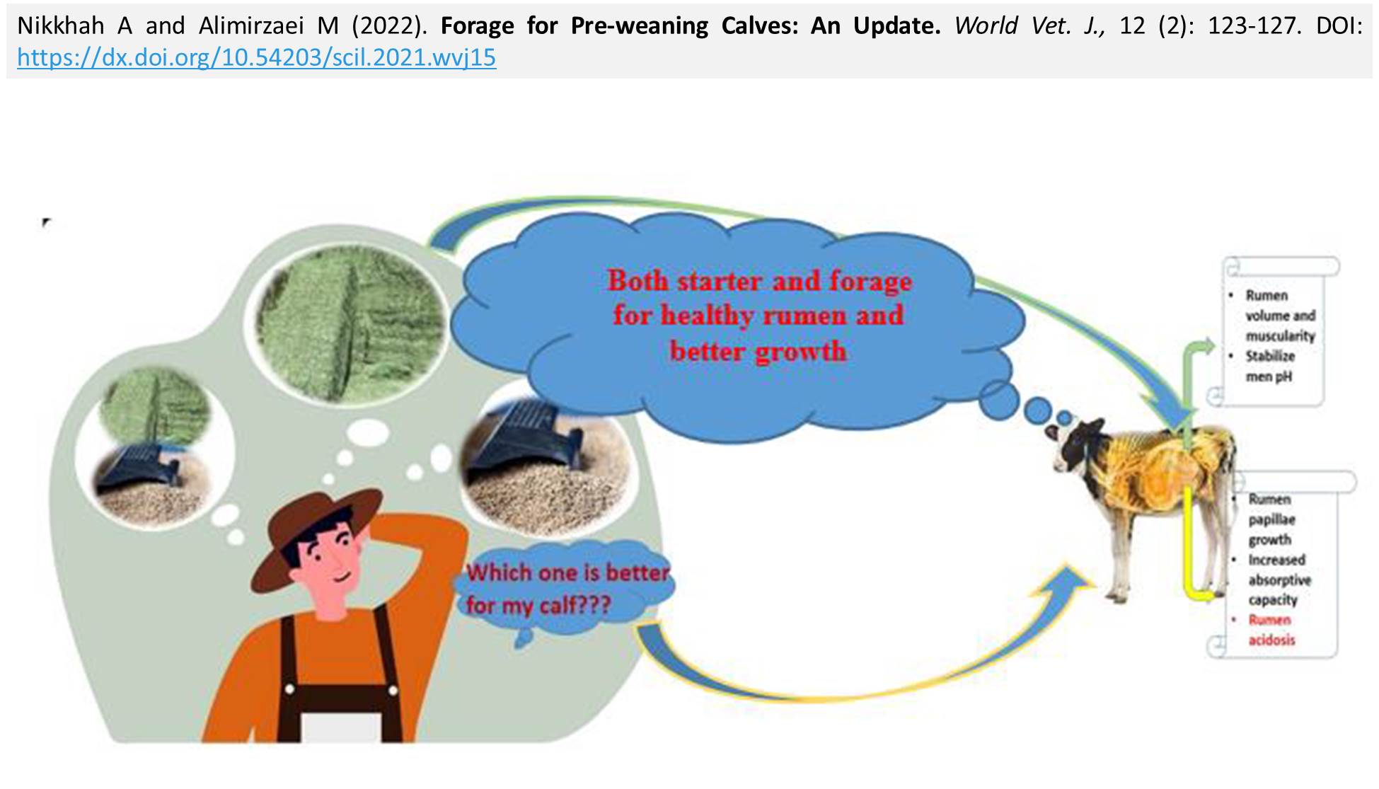
Forage for Pre-weaning Calves: An Update
Nikkhah A and Alimirzaei M.
World Vet. J. 12(2): 123-127, 2022; pii:S232245682200015-12
DOI: https://dx.doi.org/10.54203/scil.2022.wvj15
ABSTRACT: Forage nutrition for pre-weaning calves hosts numerous practical questions and on-farm challenges. The present review article aimed to update and address the biological consequences of forage provision to pre-weaned dairy calves. Health, nutrient intake (milk plus solid feed), and rumen development are the most important factors related to calf growth in pre- and post-weaning periods. A growing body of evidence suggests that the health and growth performance of dairy calves in the pre-weaning period are associated with their later performance as dairy cows. It seems that starter feeding strategies, including grain type, processing method, feed texture, and forage inclusion during the critical pre-weaning period may have profound effects on rumen function and calf performance. It is well understood that grain fermentation by-products are essential for increased growth and absorptive capacity of the rumen papillae. Forage provision as a part of a starter diet has been a topic of recent research. The rumen pH is the main factor altering the fate of fermentation and eventually animal health. In the pre-weaned calf, two major hypotheses exist regarding forage feeding. The first hypothesis describes that the rumen is not completely developed in pre-weaned calves and forage provision during this period might increase gut fill, and hence, decrease starter intake. It is believed that depressed starter intake may limit energy intake and finally suppress calf growth rate. The second hypothesis indicates that the rumen pH may decline as calves age and starter intake increases. Accordingly, forage inclusion in calf starter diets could prevent further rumen pH decline and subsequent negative consequences while improving starter intake and calf growth. Research data regarding these hypotheses are controversial. Many factors, such as milk feeding method, grain, forage type, and experimental conditions could affect calf responses to dietary forage. The current review focused on the biological consequence of forage provision to young calves to provide a practical framework for better use of forages in pre-weaned calves feeding programs.
Keywords: Forage, Growth, Pre-weaned calf, Rumen development
[Full text-PDF] [Crossref Metadata] [Scopus] [Export from ePrint] [How to Cite]
 Review
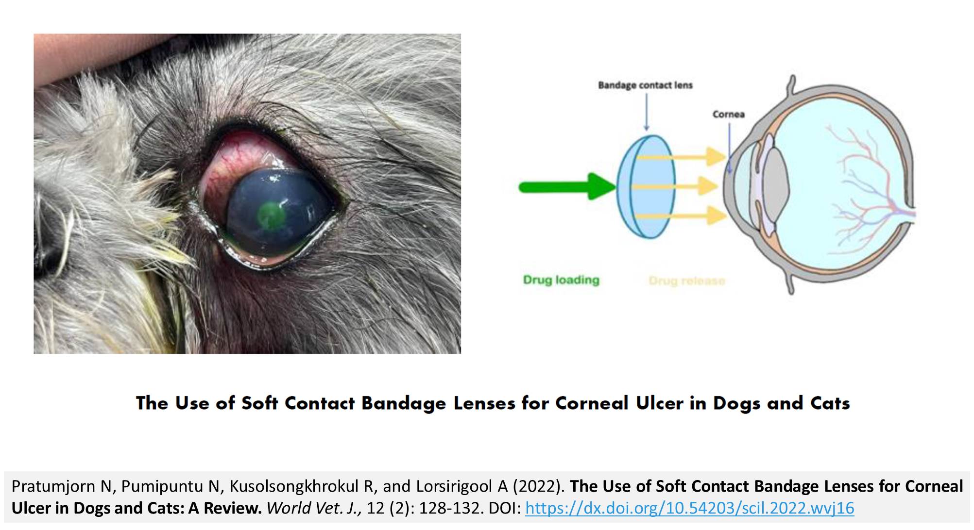
The Use of Soft Contact Bandage Lenses for Corneal Ulcer in Dogs and Cats: A Review
Pratumjorn N, Pumipuntu N, Kusolsongkhrokul R, and Lorsirigool A.
World Vet. J. 12(2): 128-132, 2022; pii:S232245682200016-12
DOI: https://dx.doi.org/10.54203/scil.2022.wvj16
ABSTRACT: A corneal ulcer is the characteristic of the destruction of the corneal epithelium layer and loss of the stroma layer at various depths. At present time, soft bandage contact lenses are used in many countries for corneal ulcers in dogs and cats to protect their cornea, increase contact time with topical eyes solutions, and support corneal reepithelialization. This article aimed to review information on the use of soft contact bandage lenses to treat corneal ulcers in dogs and cats interms of their efficacy and precaution. The results of the present review have revealed that soft contact bandage lenses are used to protect the cornea, enhance contact time with topical eye treatments, reduce median healing time, and provide comfort in dogs and cats with corneal ulcers. In the case of serious infections and dry eyes, soft contact bandage lenses are not recommended.
Keywords: Corneal ulcer, Incidence, Soft contact bandage lenses, Treatment
[Full text-PDF] [Crossref Metadata] [Scopus] [Export from ePrint] [How to Cite]
 Research Paper
The Impact of Black Soldier Fly (Hermetia illucens) as Feed Supplementation on Productive and Physiological Performance of Broiler Chickens
El-Kaiaty AM, Atta A-E-R.M, Dawa DT, and El-sayed TR.
World Vet. J. 12(2): 133-140, 2022; pii:S232245682200017-12
DOI: https://dx.doi.org/10.54203/scil.2022.wvj17
ABSTRACT: A total of 450 broiler chicks (Ross 308) were used to evaluate the effect of different inclusion levels of a partially black soldier fly (BSF), BSF Powder (BSFP), BSF Puré (BSFPr), and BSF frozen whole larvae (BSFL) on the growth performance, blood parameters, humoral immune response, and intestinal bacterial count of broiler chickens. The chickens were reared from day 1 to 35 and assigned to the control and 9 dietary groups with different forms of BSF (3 replicates per group with 15 chicks). Black soldier fly was included at levels of 2%, 4%, and 6% for BSFP, BSFPr, and BSFL, respectively, in the starter and growing diets. The results indicated similar body weight, weight gain, and the growth rate in chickens fed 4% BSFP, and 2% BSFPr during the experiment. There was a marked difference in blood parameters due to the different BSF forms and included percentages. The humoral immunity antibody titers against the Newcastle disease virus fluctuated among the experimental groups of different ages. Finally, it could be concluded that the BSF can be incorporated at a level of 4% in the form of powder and Puré in a broiler diet which seemed to be adequate to achieve the favorable results in growth performance, blood parameters, immunity, and bacteriological examination.
Keywords: Black soldier fly, Insects, Black solidier fly powder, Humoral immune response, Soybean substitution
[Full text-PDF] [Crossref Metadata] [Scopus] [Export from ePrint] [How to Cite]
 Research Paper
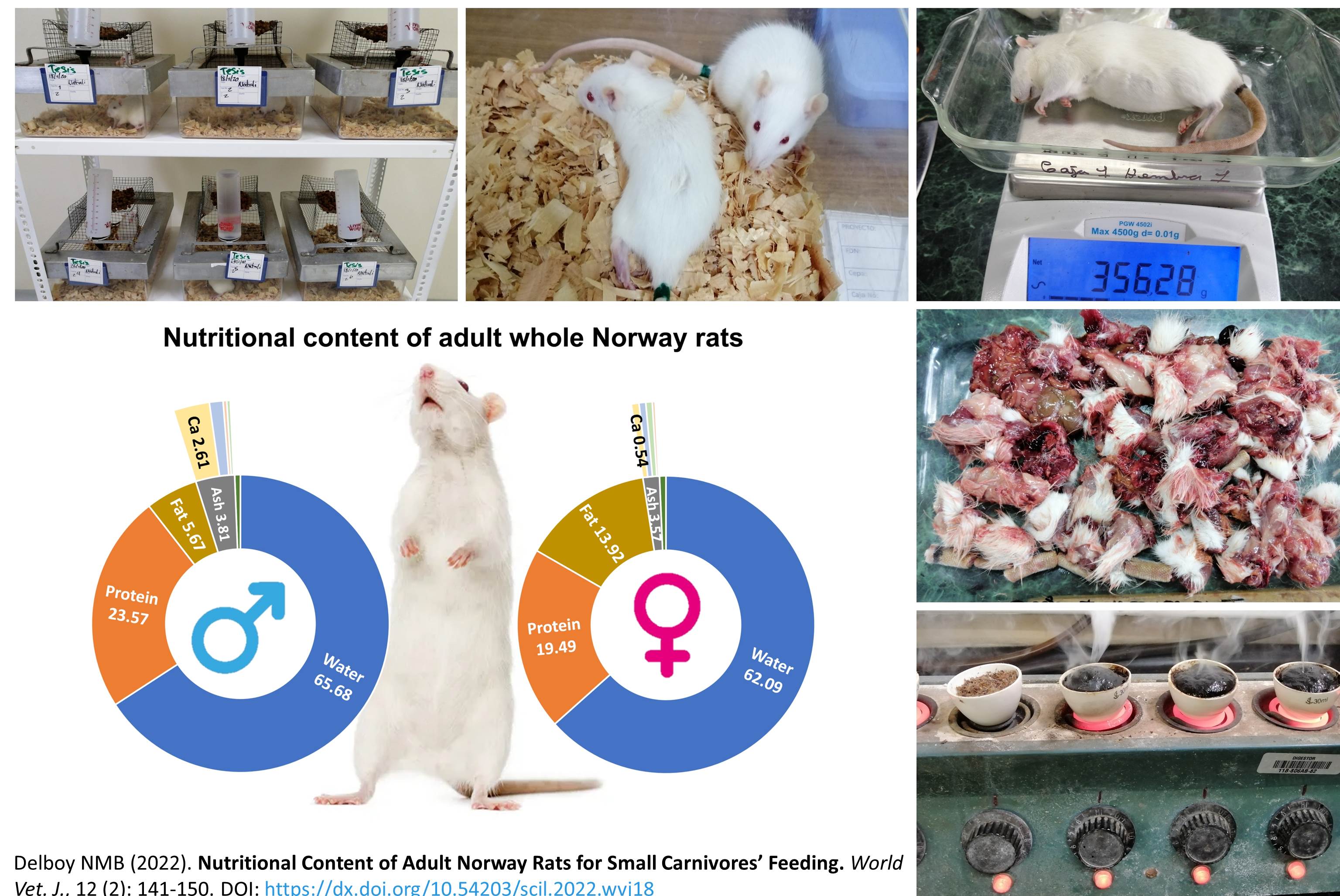
Nutritional Content of Adult Norway Rats for Small Carnivores’ Feeding
Delboy NMB.
World Vet. J. 12(2): 141-150, 2022; pii:S232245682200018-12
DOI: https://dx.doi.org/10.54203/scil.2022.wvj18
ABSTRACT: Rats are the natural diet of many free-ranging carnivores. They are also fed to small carnivore mammals, raptor birds, and reptiles in captivity as a sole or partial diet, however, little is known about the nutrients that a rat can provide as animal feed. This study aimed to determine the nutritional content of the whole captive-bred Norway rats. A total of 12 randomly selected weaned male and female Wistar Norway rats were fed ad libitum with a local dry dog food diet. The rats were weighed weekly until an average weight of 300 g was reached. Biochemical and mineral analyses were carried out for each rat. The results of the study showed significant differences between male and female rats in terms of growth rate, crude protein, total fat, and calcium concentrations. Males presented a faster growth rate and reached the desired weight in around half the time (6 weeks), compared to females (13 weeks). Moreover, males had a higher percentage of crude protein (23.57%) on a fed matter basis, calcium (2.61%), and phosphorus (0.98%). Females showed higher total fat (13.92%) and lower crude protein (19.49%), calcium (0.54%), and phosphorus (0.47%), compared to males. The results of this research may be used to determine whether a whole rat can provide all the necessary nutrients to carnivore animals commonly kept in captivity. Present findings indicated that rats could provide the necessary nutrients, however, if given as a sole diet, they could not be enough to supply the nutritional requirements of animals in the long term.
Keywords: Carnivore nutrition, Norway rats, Nutritional Content, Wistar rat
[Full text-PDF] [Crossref Metadata] [Scopus] [Export from ePrint] [How to Cite]
 Research Paper
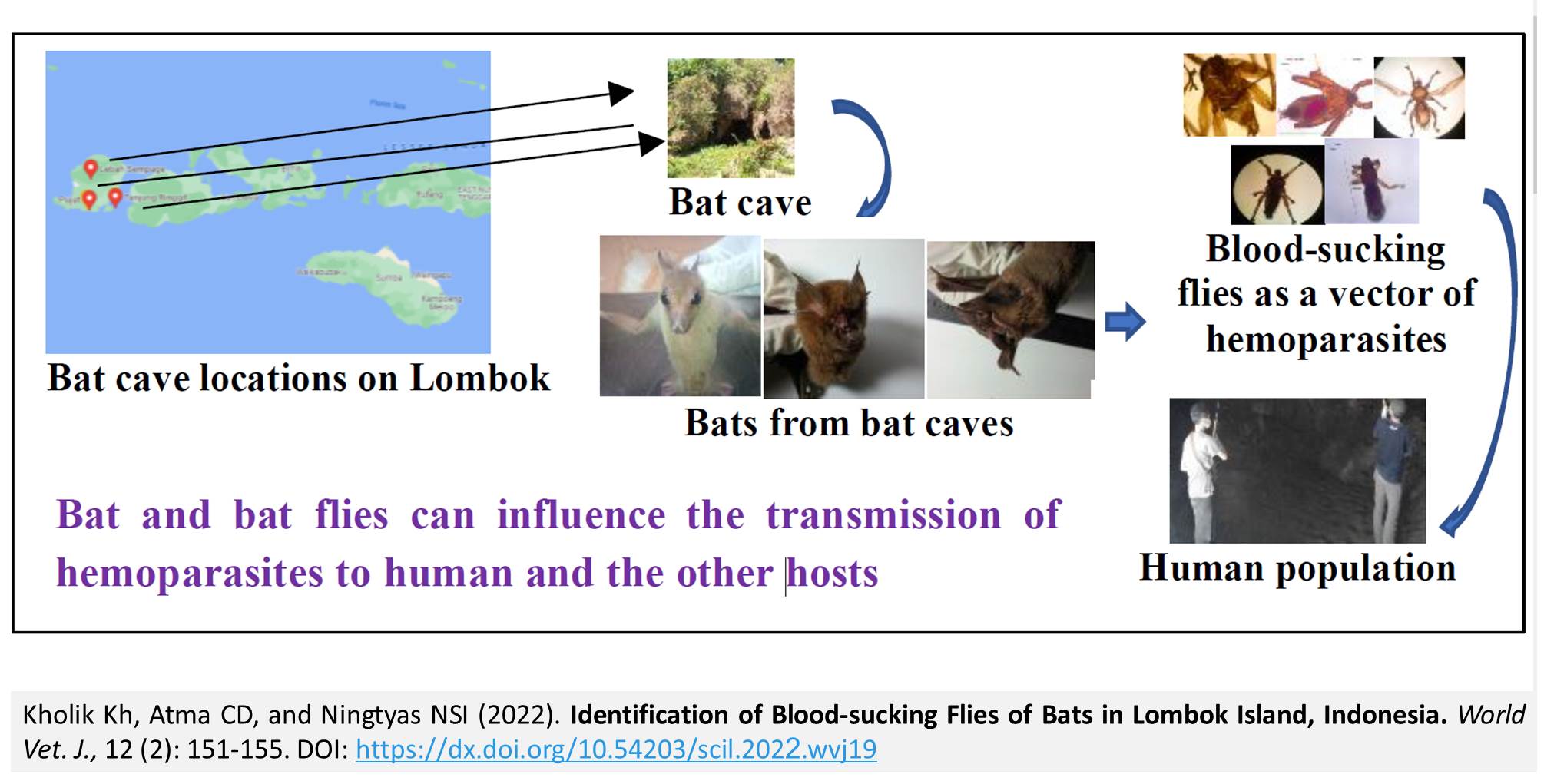
Identification of Blood-sucking Flies of Bats in Lombok Island, Indonesia
Kholik Kh, Atma CD, and Ningtyas NSI.
World Vet. J. 12(2): 151-155, 2022; pii:S232245682200019-12
DOI: https://dx.doi.org/10.54203/scil.2022.wvj19
ABSTRACT: Bats and blood-sucking bat flies have an important role in transmitting several hemoparasites. Bat flies have been identified as vectors transmitting hemoparasites from wild bats. The purpose of the present study was to identify bats and their blood-sucking flies as vectors of hemoparasites in bat caves located at Lombok Island, Indonesia. In the course of the study, a survey was conducted on three bat caves from September to December 2018. The bats were captured by a net trap and the species of bats and bat flies were identified. A total of 66 captured bats were identified as Hipposideros species (n = 28), Eonycteris spelaea (n = 23), and Taphozouss species (n = 15). The blood-sucking flies were identified as Eucampsipoda sundaica on Eonycteris spelaea, and Stylidia cf. euxesta, Brachytarsina species, Raymondia species, and Megastrebla nigriceps on Hipposideros species. The results showed that five species of blood-sucking flies were present in captured bats. The bat and blood-sucking flies can influence the transmission of Polychromophilus species, Babesia species, Plasmodium species, and Trypanosoma species to humans and other hosts.
Keywords: Bats, Blood-sucking flies, Hemoparasites, Lombok, Vector
[Full text-PDF] [Crossref Metadata] [Scopus] [Export from ePrint] [How to Cite]
 Research Paper
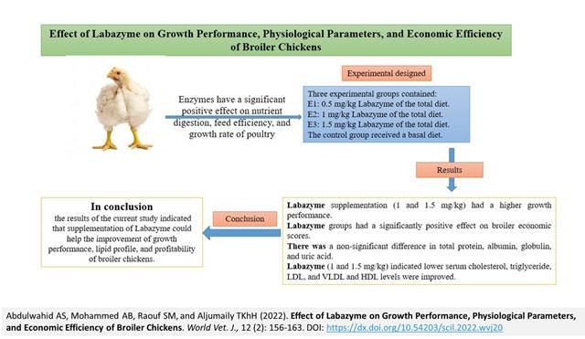
Effect of Labazyme on Growth Performance, Physiological Parameters, and Economic Efficiency of Broiler Chickens
Abdulwahid AS, Mohammed AB, Raouf SM, and Aljumaily TKhH.
World Vet. J. 12(2): 156-163, 2022; pii:S232245682200020-12
DOI: https://dx.doi.org/10.54203/scil.2022.wvj20
ABSTRACT: Enzymes have a significant positive effect on nutrient digestion, feed efficiency, and growth rate of poultry. The current experiment aimed to determine the optimal dosage levels of Labazyme as feed additives. A total of 240 one-day-old broiler chickens (Ross 308) were randomly assigned to four groups with three replicates. The feeding experiment was carried out from hatching to day 42 of age. Three experimental groups contained Labazyme at 0.5, 1, and 1.5 mg/kg of the total diet. The control group received a basal diet. Growth performance, European performance efficiency index (EPEI), production index (PI), biochemical and lipid profiles, as well as antioxidant parameters were then measured. The results showed that chickens fed Labazyme supplementation (1 and 1.5 mg/kg) had a higher growth performance than those in the control group. Nonetheless, there was a significant difference between the Labazyme and the control group in terms of feed intake. In addition, Labazyme groups had a significantly positive effect on broiler economic scores. The EPEI and PI of the Labazyme-fed chickens were both higher than the control. There was a non-significant difference in total protein, albumin, globulin, and uric acid. The serum glucose level of the chickens fed Labazyme (1 and 1.5 mg/kg) was lower, compared to the control group. In contrast, chickens that consumed a diet supplemented with Labazyme 1 and 1.5 mg/kg indicated lower serum cholesterol, triglyceride, low-density lipoprotein, and very-low-density lipoprotein levels in broilers, compared to the control group. Serum high-density lipoprotein levels were improved and more pronounced in chickens fed Labazyme, compared to the control group. In conclusion, the results of the current study indicated that supplementation of Labazyme could help the improvement of growth performance, lipid profile, and profitability of broiler chickens.
Keywords: Broiler, Labazyme, Lipid profiles, Production index
[Full text-PDF] [Crossref Metadata] [Scopus] [Export from ePrint] [How to Cite]
 Research Paper
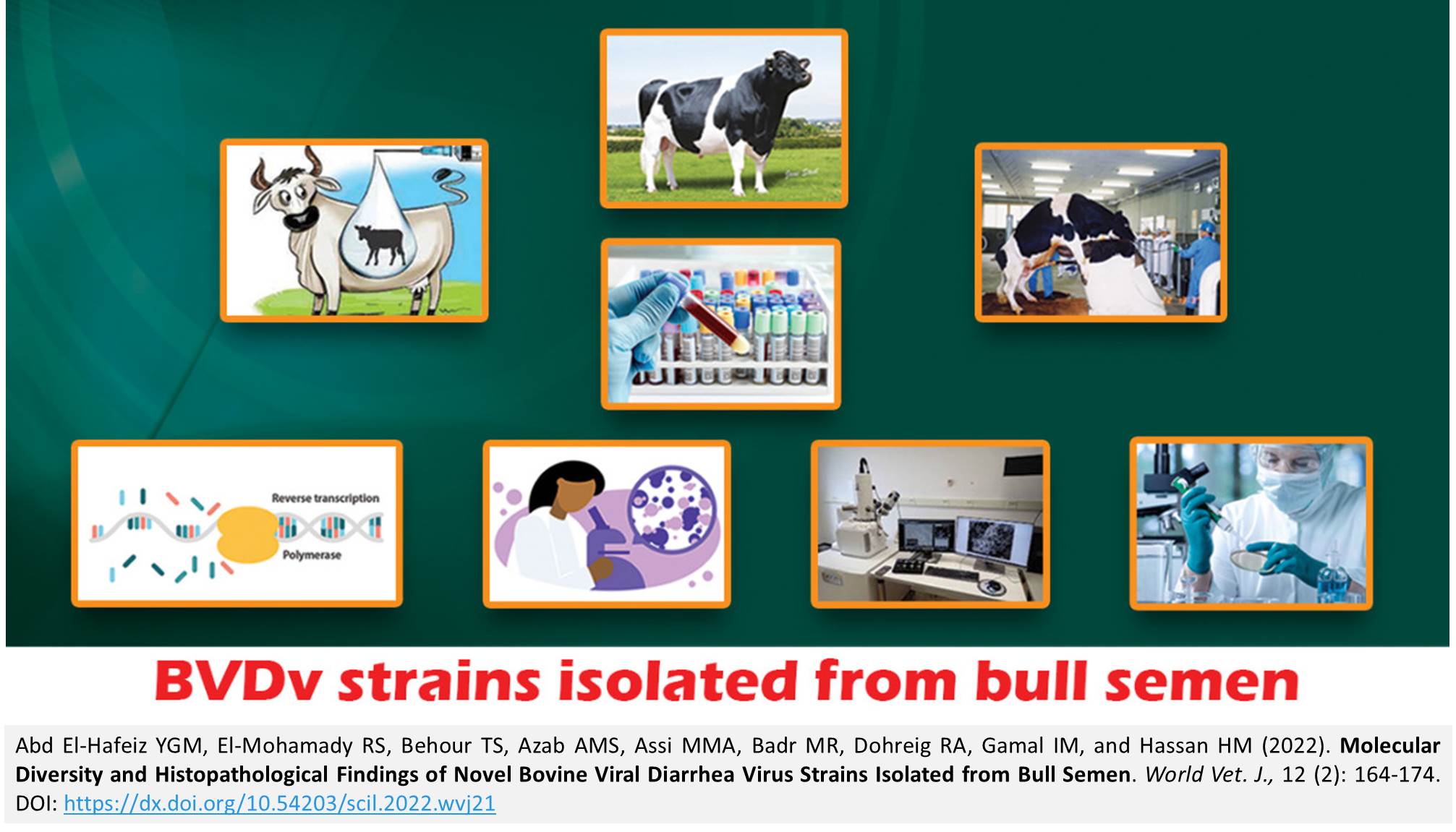
Molecular Diversity and Histopathological Findings of Novel Bovine Viral Diarrhea Virus Strains Isolated from Bull Semen
Abd El-Hafeiz YGM, El-Mohamady RS, Behour TS, Azab AMS, Assi MMA, Badr MR, Dohreig RA, Gamal IM, and Hassan HM.
World Vet. J. 12(2): 164-174, 2022; pii:S232245682200021-12
DOI: https://dx.doi.org/10.54203/scil.2022.wvj21
ABSTRACT: Bovine viral diarrhea virus (BVDV) is one of the most common viral pathogens affecting the cattle industry worldwide. The present study aimed to molecularly characterize BVDV isolates that are currently circulating in breeding bulls farmed with cattle suffering from reproductive disorders, and also to assess the consequences of BVDV infection on bulls’ semen quality and conception, and its pathological effects on the structure of testicular tissue and spermatozoa. For this purpose, semen, serum, and testicular samples were collected from four breeding bulls in four private dairy farms in the governorates of Kafr-El Sheik, Beni-Suef, Giza, and Assuit, in Egypt from April 2019 to May 2020. An evaluation of sperm abnormalities was carried out by assessing the integrity of the plasma and acrosomal membranes where severe damage and abnormalities were found. Ultrastructure analysis of the spermatozoa by transmission electron microscopy revealed the presence of a swollen plasma membrane with segmented outer acrosomal membrane of spermatozoa and vacuolar degenerated mitochondria. Histopathological examination of testicular and epididymal tissues indicated moderate to severe degenerative effects of virus infection on seminiferous tubules with hypospermatogenesis. By detection of virus antigen in the serum samples using ELISA, bulls were identified as persistently infected with BVDV. Virus isolation revealed four noncytopathic (NCP-BVDV) strains that were confirmed by fluorescent antibody technique (FAT) and amplification of the 5′ untranslated genomic region (5’UTR) and molecularly typed by amplification of the Erns glycoprotein region. Isolates’ Phylogenetic analysis revealed two subgenotypes: BVDV-1b (Genbank accession numbers; LC634512, LC634513, LC634515) and BVDV-1d (LC634516). According to the knowledge of the authors of the present study, the circulation of the BVDV-1d subgenotype is not reported in Egypt. Therefore, it would be of great importance to track circulating strains in specific countries for successful vaccination programs or accurate diagnostic tests, and this necessitates regular updates.
Keywords: BVDV, Isolation, Spermatozoa ultrastructure, Sperm abnormalities, Testicular histopathology
[Full text-PDF] [Crossref Metadata] [Scopus] [Export from ePrint] [How to Cite]
 Research Paper
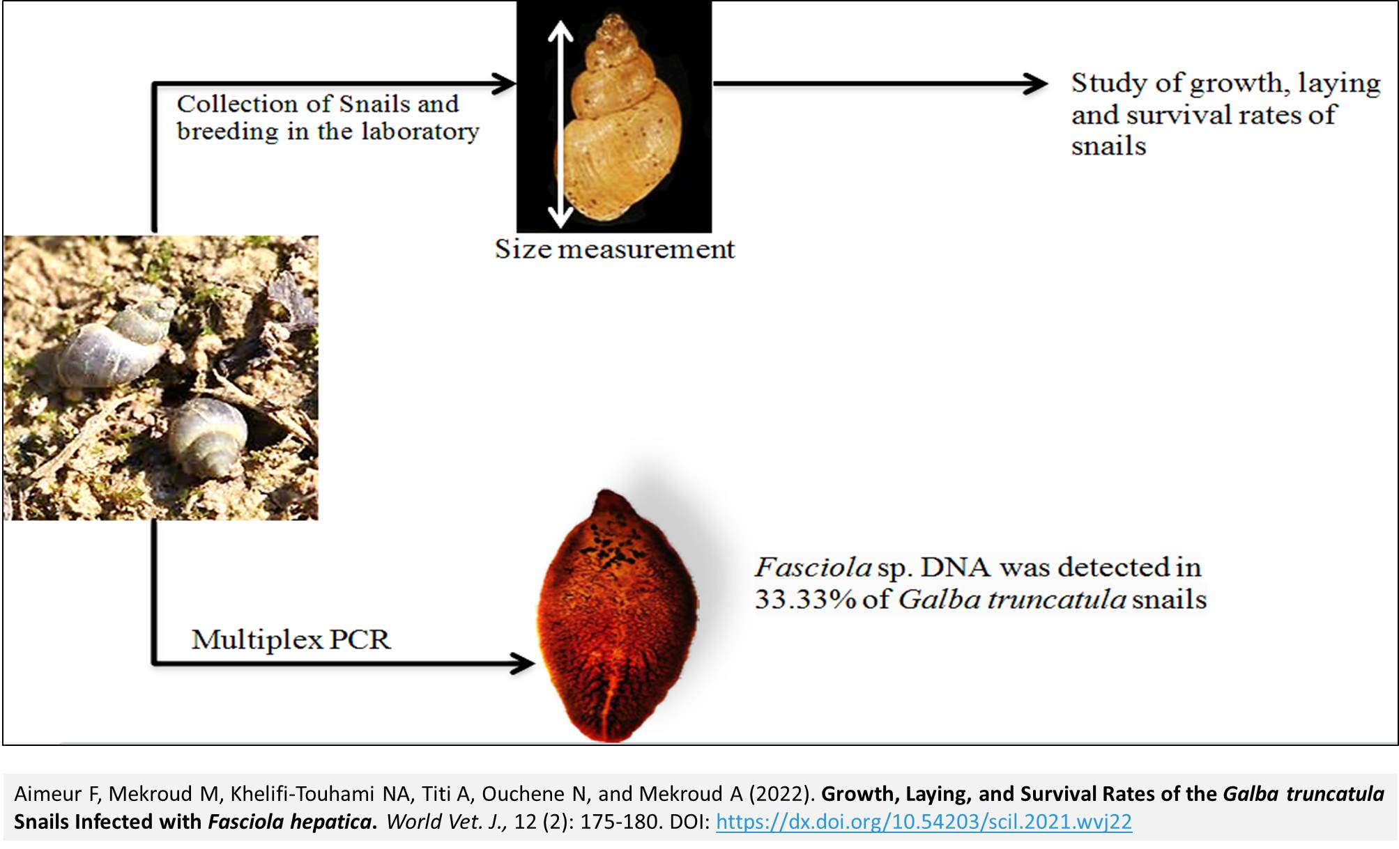
Growth, Laying, and Survival Rates of the Galba truncatula Snails Infected with Fasciola hepatica
Aimeur F, Mekroud M, Khelifi-Touhami NA, Titi A, Ouchene N, and Mekroud A.
World Vet. J. 12(2): 175-180, 2022; pii:S232245682200022-12
DOI: https://dx.doi.org/10.54203/scil.2022.wvj22
ABSTRACT: Fasciolosis is one of the most important parasitic diseases in ruminants in Algeria, of which the intermediate host is Galba truncatula (G. truncatula) snail. The current study aimed to investigate the prevalence of Fasciola sp. in naturally infected G. truncatula snails using multiplex PCR. Secondly, it was targeted toward examining the rate of growth, survival, and laying of the snails in experimental conditions during 6 weeks of rearing in three tanks. This study was conducted in two different regions of Algeria, namely El Tarf and Constantine. The investigated tanks 1, 2, and 3 consisted of 12 (size 3-4 mm), 30 (size 5-6 mm), and 30 (size 7-8 mm) snails, respectively. Fasciola sp. DNA was detected in 33.33% of G. truncatula snails (25% in Constantine and 42.85% in El Tarf). The total survival rates in the first, second, and third tanks were 50%, 43.3%, and 40%, respectively. The obtained results indicated that the growth rate of the snail depended on its initial size (the smaller the initial size, the higher the weekly growth rate). The total growth rates were 3, 1.7, and 1.1 mm in tanks 1, 2, and 3, respectively. The use of multiplex PCR indicated a relatively high level of infestation of the snails by Fasciola sp. Snails larger than 7 mm had the highest lay rate. Further studies are needed to investigate other snails that may be infested with Fasciola sp.
Keywords: Fasciola, Galba truncatula, PCR multiplex, Snail
[Full text-PDF] [Crossref Metadata] [Scopus] [Export from ePrint] [How to Cite]
 Short Communication
Parascaris equorum in Horses of Payakumbuh City, West Sumatra, Indonesia
Zelpina E, Sujatmiko S, Silvia Noor P, and Lefiana D.
World Vet. J. 12(2): 181-185, 2022; pii:S232245682200023-12
DOI: https://dx.doi.org/10.54203/scil.2022.wvj23
ABSTRACT: Parascaris equorum is a species of the Ascarididae family which belongs to the phylum nematoda (roundworms) and is a type of parasite that affects equine health, performance, and production. The current study was carried out from April to August 2021 to determine the prevalence of equine Parascaris equorum in horses residing in Payakumbuh City, Indonesia. A total number of 128 fecal samples from horses were examined in the current study. Coprological examination was performed for the detection of Parascaris equorum eggs inside the amassed samples. The overall occurrence of Parascaris equorum was 14.06% (18 out of 128). The prevalence rates of sub-districts differed with the highest in East at 18.18% (8/44), followed by North, South, and West at 16.66% (6/36), 14.06% (2/22), and 7.7% (2/26), respectively. The obtained results indicated a significant difference in the prevalence rate of Parascaris equorum between males and females as well as those horses aged < 5 years (26.22%) and > 5 years (3%). Therefore, it is critical to not only enhance horse health management, maintenance, and health but also to provide anti-parasitic medications on a regular basis.
Keywords: Gastrointestinal nematode, Horse, Parascaris equorum, Prevalence
[Full text-PDF] [Crossref Metadata] [Scopus] [Export from ePrint] [How to Cite]
 Research Paper
Antimicrobial Resistance and Virulence Genes of Campylobacter jejuni Isolates from Diarrheic Sheep
Hafez A.
World Vet. J. 12(2): 186-196, 2022; pii:S232245682200024-12
DOI: https://dx.doi.org/10.54203/scil.2022.wvj24
ABSTRACT: One of the important agents causing gastroenteritis worldwide is Campylobacter jejuni (C. jejuni). The current study aimed to detect five virulence genes (flaA, virB11, ciaB, iam, and dnaJ) and two antibiotic resistance genes (gyrA and tetO) in C. jejuni obtained from sheep stool. The virulence genes were detected by PCR in 64 C. jejuni strains. The phenotypic resistance to five selected antibiotics (Ciprofloxacin, Erythromycin, Gentamycin, Streptomycin, and Tetracycline) was screened with the microdilution method. The isolates with antibiograms were tested for detection of gyrA and tetO genes via PCR using specific primers. The virulence genes flaA (32%) and dnaJ (29%) had the highest prevalence. The tested isolates of C. jejuni revealed high resistance to both quinolone (68.3%) and tetracycline groups (48.4%) with an increased prevalence of antibiotic resistance of gyrA and tetO genes. Gentamycin and erythromycin offered better alternative drugs for the treatment of campylobacteriosis. To generalize the findings, extensive profiling that involves more virulence genes is required in several strains of Campylobacter.
Keywords: Antibiotic resistance, Campylobacter jejuni, Sheep, Virulence genes
[Full text-PDF] [Crossref Metadata] [Scopus] [Export from ePrint] [How to Cite]
 Research Paper
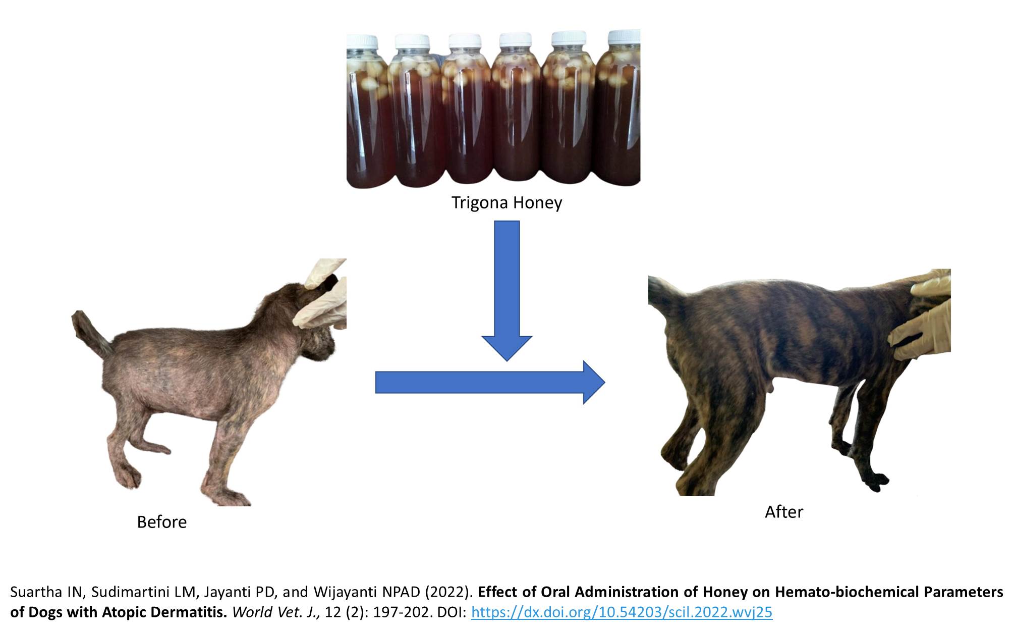
Effect of Oral Administration of Honey on Hemato-biochemical Parameters of Dogs with Atopic Dermatitis
Suartha IN, Sudimartini LM, Jayanti PD, and Wijayanti NPAD.
World Vet. J. 12(2): 197-202, 2022; pii:S232245682200025-12
DOI: https://dx.doi.org/10.54203/scil.2022.wvj25
ABSTRACT: Honey from Trigona species is widely used as herbal medicine in humans due to its antimicrobial, anti-inflammatory, and antioxidant effects as well as the potential to increase body resistance and boost blood formation. The current study aimed to determine the hemato-biochemical profile of dogs with atopic dermatitis treated with Trigona honey. The hematology profile included the measurement of erythrocytes, hemoglobin, hematocrit, and erythrocyte index, as well as blood biochemical parameters, including aspartate aminotransferase (AST), alanine transaminase (ALT), and blood sugar. A total of 12 local dogs aged 4 months old were divided into two treatment groups, namely the control group (G1) and treatment with liquid Trigona honey at a dosage of 5 ml/dog/day (G2) for 35 days. Then, blood was collected and tested for routine and chemical blood assay. The results showed that the administration of fresh Trigona honey (5 ml/day for 5 weeks) exhibited a significant increase in most of hematological variables of dogs with atopic dermatitis, compared to G1. The results of blood biochemical profiles (AST, ALT, and blood glucose) remained unaffected by the treatment of Trigona honey. It can be concluded that honey from Trigona spp. was safe to be given to the dogs with dermatitis and no adverse physiological effects were observed during the present study.
Keywords: Blood, Dermatitis, Dog, Hemato-biochemical, Trigona species honey
[Full text-PDF] [Crossref Metadata] [Scopus] [Export from ePrint] [How to Cite]
 Research Paper
The Transmission Pattern of Amoebiasis in Bale Zone, South East Ethiopia
Jilo SA, Kadir MA, Hussein JA, and Nair SP.
World Vet. J. 12(2): 203-213, 2022; pii:S232245682200026-12
DOI: https://dx.doi.org/10.54203/scil.2022.wvj26
ABSTRACT: Amoebiasis is a primarily zoonotic disease, mainly transferred through the fecal-oral route and waterborne. Amoebiasis is still a big challenge for human and animal health and is a major cause of diarrhea in developing countries, including Ethiopia. Therefore, the study was conducted to assess the epidemiology of the disease in humans, dogs, and the occurrence of the parasite in water bodies. A prospective cross-sectional study was conducted in selected districts of the Bale zone in southeastern Ethiopia. Pet owners were selected randomly. Socio-demographic data were collected using a questionnaire and fecal samples were used to perform microscopic examination. A total of 383 fecal samples of humans, 383 fecal samples of dogs, and 58 water samples were studied from December 2019 to July 2020. Of 383 humans, 179 were males and 186 were females, while 94 individuals were grouped as children younger than 8 years, 164 were grouped as youth within the age range of 8-18 years, and 125 were grouped as adults who were older than 18 years. Of 383 local breeds, dogs were grouped as 87 puppies younger than one year, 192 young dogs with the age range of 1-2 years, and 104 adult dogs who were older than 2 years. Fecal samples were taken from 173 male and 210 female dogs. The water samples were taken randomly from the water sources (river, lake, pond, or water tank) at different sites where dogs and humans can easily contact water to use for different purposes. Of the total samples, 70 humans (18.3%), 63 dogs (16.5%), and 16 water samples (27.6%) were contaminated with the parasite. The major risk factors for the transmissions of parasites were contaminated drinking water, large family size, open-air defecation, and improper handwashing. The present study revealed that the human reservoir was a major risk factor for the spread and transmission of amoebiasis in dogs. The high prevalence of the disease might be due to open-air defecation, unhygienic health practices, domestic animals inside the houses, and using local water bodies as a drinking source.
Keywords: Amoeba, Dog, Human, Transmission, Water
[Full text-PDF] [Crossref Metadata] [Scopus] [Export from ePrint] [How to Cite]
 Review
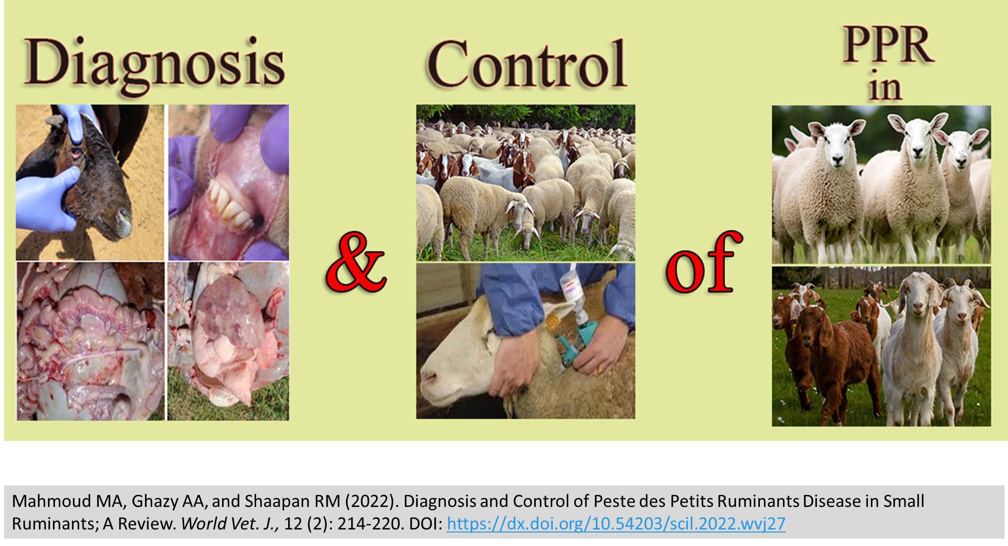
Diagnosis and Control of Peste des Petits Ruminants Disease in Small Ruminants; A Review
Mahmoud MA, Ghazy AA, and Shaapan RM.
World Vet. J. 12(2): 214-220, 2022; pii:S232245682200027-12
DOI: https://dx.doi.org/10.54203/scil.2022.wvj27
ABSTRACT: Peste des Petits Ruminants (PPR) is an acute highly contagious febrile disease of sheep and goats characterized by erosive and necrotizing stomatitis and associated with severe pneumo-enteritis and bronchopneumonia ended by recovery or death. The aim of the present study was to throw light on the diagnosis and control of PPR. Diagnosis of PPR depends on clinical signs, pathological lesions, and specific detection of the viral antigen, viral genome, or specific antibodies by serological tests and nucleic acid-based assays. The most commonly used diagnostic techniques are cell culture isolation, agar gel immunodiffusion, hemagglutination tests, immunocapture ELISA, and competitive ELISA. In addition to the abovementioned techniques, virus neutralization tests and reverse transcriptase PCR are used. Peste des Petits Ruminants is characterized by high fever associated with watery nasal and ocular discharges, mucopurulent stomatitis, and broncho-pneumonia. Moreover, severe bloody diarrhea and the disease associated with high levels of mortality reached up to 90%. The diagnosis of viral diseases is important in determining the control strategies. Therefore, it can be concluded that recent diagnostic tools are urgently needed not only for the diagnosis but also for following-up combating programs and control of viral diseases. Early and rapid complete identification of infectious viral agents in small ruminants as well as in the surrounding environment is recommended for effective control of PPR. The control program depends mainly on vaccination, hygiene and sanitation measures, and effective quarantine measures.
Keywords: Control, Dairy Ruminants, Diagnosis, Goat, Peste des Petits, Sheep
[Full text-PDF] [Crossref Metadata] [Scopus] [Export from ePrint] [How to Cite]
 Review
Bats and Antibiotic Resistance: A Culprit or a Victim?
Garcês A.
World Vet. J. 12(2): 221-229, 2022; pii:S232245682200028-12
DOI: https://dx.doi.org/10.54203/scil.2022.wvj28
ABSTRACT: In the last decades, the increase of antimicrobial resistance bacteria has become a concern for public health. Bats’ ability to fly, form colonies for a long lifespan, and inhabit a variety of diverse ecological niches make them successful species in terms of adaptation and distribution on earth. Moreover, these characteristics let them act as the potential natural reservoir of numerous zoonotic pathogens (bacteria, viruses, fungi). Bat bacteriome knowledge is still very scarce, but a few studies have indicated that bats are hosts of antimicrobial resistance and play an important role in the dispersion of resistance in the environment. Moreover, bats are vulnerable to acquiring these pathogens since they sometimes live in close contact with humans and domestic animals. Therefore, the present study aimed to compile the latest studies that describe the presence of antibiotic-resistant in bats. Based on the papers analyzed for this review, it is possible to conclude that bats are hosts of pathogenic bacteria that carry numerous antibiotic resistance. Extended-spectrum b-lactamases (ESBLs) or Methicillin-resistant Staphylococcus which nowadays days are a great public health concern, have already been reported in these animals, with some isolated strains being of Human origin. Although not completely understood regarding the dynamics and transmission routes, bats seem to have an important role in the dissemination and acquisition of antibiotic resistance in the environment. They can be contaminated by bacteria with antibiotic resistance and disperse through the environment. However, they also can be the host of bacteria that carry antibiotic resistance.
Keywords: Antibiotic-Resistant, Bacteria, Bat, Chiropters, Zoonoses
[Full text-PDF] [Crossref Metadata] [Scopus] [Export from ePrint] [How to Cite]
Previous issue | Next issue | Archive
![]() This work is licensed under a Creative Commons Attribution 4.0 International License (CC BY 4.0).
This work is licensed under a Creative Commons Attribution 4.0 International License (CC BY 4.0).