Previous issue | Next issue | Archive
![]() Volume 12 (3); September 25, 2022 [Booklet] [EndNote XML for Agris]
Volume 12 (3); September 25, 2022 [Booklet] [EndNote XML for Agris]
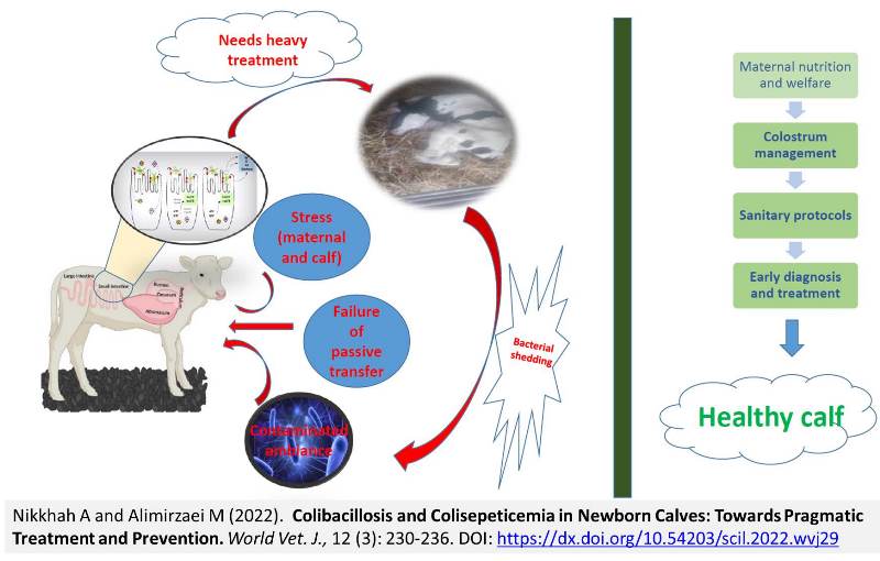
Colibacillosis and Colisepeticemia in Newborn Calves: Towards Pragmatic Treatment and Prevention
Nikkhah A and Alimirzaei M
World Vet. J. 12(3): 230-236, 2022; pii:S232245682200029-12
DOI: https://dx.doi.org/10.54203/scil.2022.wvj29
ABSTRACT: Diarrhea is the most perturbing disease in dairy and beef industries worldwide, leading to significant rates of morbidity and mortality as well as economic losses. The objective of this review article was to delineate the pathophysiology and practical biology of colisepticemia in neonatal calves. Preventive and therapeutic protocols were also presented and discussed from a new integrative perspective. Notably, the situation can be the most deleterious in case diarrhea turns into septicemia. Under such circumstances, the mortality rate may be remarkably high and hard to control. Escherichia coli (E. coli) is an invasive and opportunistic bacteria causing severe diarrhea (colibacillosis) and colisepticemia in newborn calves. Colisepticemia is commonly prevalent in 2-5 days old calves, and colostral immunity is considered the first defensive line against E. coli infection. In addition to colostrum feeding quality and management, other management factors, such as dry cow nutrition and welfare, newborn calf welfare and nutrition, housing system, sanitation protocols, as well as early identification and treatment of sick calves, are important in preventing colisepticemia. In conclusion, understanding the mechanism of action and transmission routes of pathogenic E. coli will provide scientific and practical insight to plan preventive and therapeutic protocols decisively and successfully.
Keywords: Diarrhea, Mortality, Newborn calf, Pragmatic Prevention, Septicemia
[Full text-PDF] [Crossref Metadata] [Scopus] [Export from ePrint] [How to Cite]
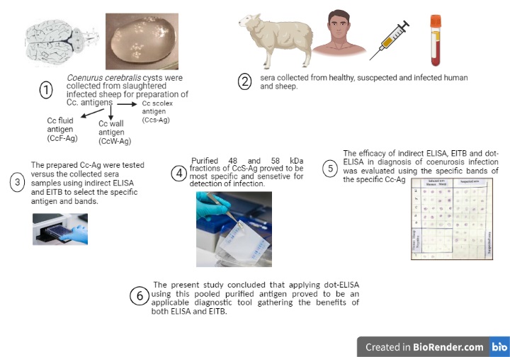
Improved Dot-ELISA Assay Using Purified Sheep Coenurus cerebralis Antigenic Fractions for the Diagnosis of Zoonotic Coenurosis
El Akkad DMH, Ramadan RM, Auda HM, Abd El-Hafez YN, El-Bahy MM, and Abdel-Radi S.
World Vet. J. 12(3): 237-249, 2022; pii:S232245682200030-12
DOI: https://dx.doi.org/10.54203/scil.2022.wvj30
ABSTRACT: Clinicians face significant problems in the diagnosis of zoonotic coenurosis. The current study aimed to develop an improved dot-Enzyme-linked-immunosorbent assay (dot-ELISA) for the diagnosis of zoonotic coenurosis using sheep Coenurus cerebralis scolices purified antigen (CcS-Ag) and to compare the obtained results with those of indirect ELISA and Enzyme-linked immunoelectrotransfer blot technique (EITB). Sera were collected from humans and sheep infected or suspected of infection with Coenurus cerebralis, control cases, and cases infected with other parasites. The CcS-Ag was proved to be the most specific antigen. This antigen was fractionated, and its specific polypeptides against anti-C. cerebralis antibodies (ACc-Ab) were identified using EITB. Fractions at the molecular weight (MW) of 48 and 58 kDa were proved as the only specific ones, eluted from the gel and concentrated, then dotted on the NC sheet as pooled antigen before its evaluation in the diagnosis of infection using dot-ELISA. Dot-ELISA demonstrated absolute 100% sensitivity and 100% specificity as recorded by EITB, compared to both fractions on a nitrocellulose (NC) sheet using surgically proved infected human or sheep sera as a gold standard. Diagnosis by ELISA using crude CcS-Ag revealed similar sensitivity but lower specificity (75%). The diagnostic accuracy of dot-ELISA was proved by comparing its results with postmortem data obtained post slaughtering of 20 suspected sheep and patients investigated by computed tomography (CT) and magnetic resonance imaging (MRI). In conclusion, the selection of specific fractions after EITB to be used in dot-ELISA improved the diagnostic value of the test as a diagnostic tool gathering the benefits of ELISA and EITB.
Keywords: Antigen,Coenurus cerebralis, Dot-ELISA, Human, Sheep, Scolices
[Full text-PDF] [Crossref Metadata] [Scopus] [Export from ePrint] [How to Cite]
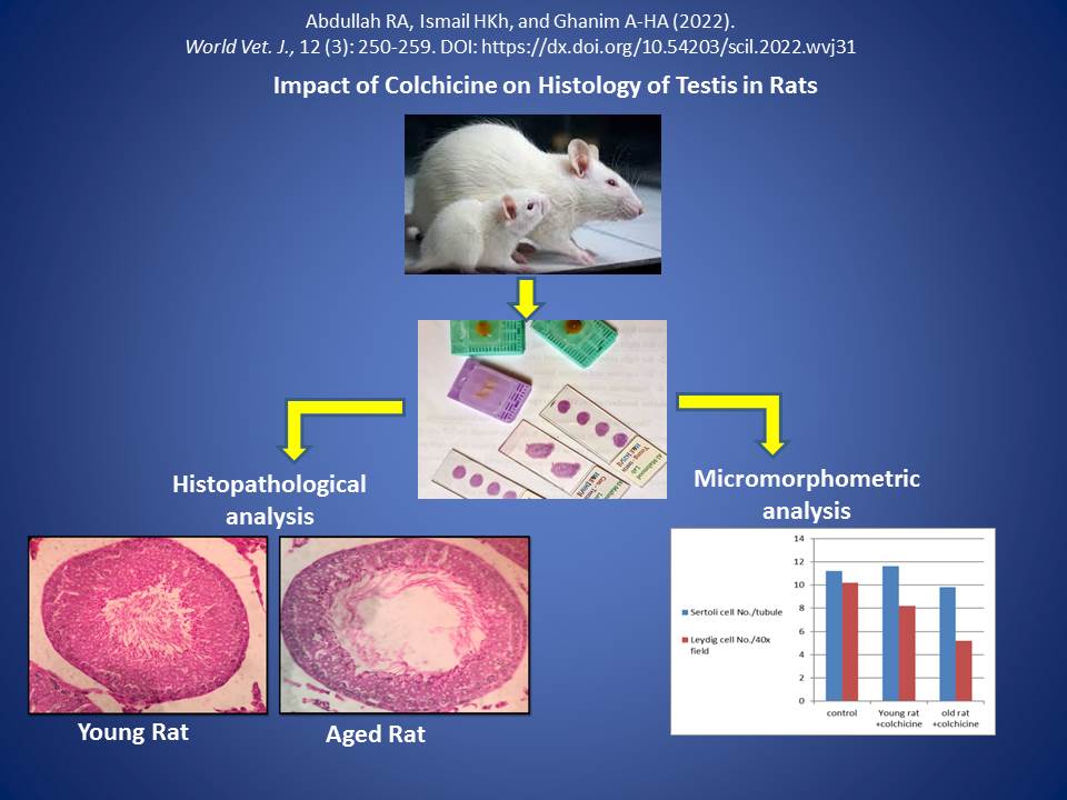
Impact of Colchicine on Histology of Testis in Rats
Abdullah RA, Ismail HKh, and Ghanim A-HA.
World Vet. J. 12(3): 250-259, 2022; pii:S232245682200031-12
DOI: https://dx.doi.org/10.54203/scil.2022.wvj31
ABSTRACT: Colchicine is a drug widely used for the management of many disorders, such as acute gout and Behçet’s disease. It is also prescribed for the treatment of pericarditis, atrial fibrillation coronary artery diseases, and secondary amyloidosis. In case this drug is used at the early stages of coronavirus infection, its anti-inflammatory properties may reduce the severe inflammatory reactions related to a cytokine storm by affecting the inflammasome. The purpose of the present study was to determine the toxicity of Colchicine on testis in rats from different age groups for 10 days. A total of 27 male Wistar rats were divided into three groups. The rats in group I (control group) were administered distilled water by oral gavage. Group II consisted of young rats (5-6 months old) who orally received Colchicine 3 mg/kg body weight. Group III entailed rats of 14-16 months who were orally administered colchicine 3 mg/kg body weight. The testis of the treated groups was dissected and examined for histological changes and morphometrical analysis. The obtained results indicated that high doses of Colchicine (3 mg/kg body weight) could induce tissue damage to the testis, including degeneration and necrosis of both Sertoli and Leydig cells with irregular divisions of germinal epithelium, even when it was used for short periods (10 days). In the elderly treated rats, there were severe tissue damages, including degeneration and necrosis of germinal epithelium with irregular divisions of germ cells, necrosis of Sertoli and Leydig cells with sloughing of germinal epithelium toward the lumen of the tubule. Therefore, there is a need to conduct more studies to investigate the side effect of Colchicine as it is excessively used in the management of coronavirus.
Keywords: Colchicine, Histology, Morphometric trait, Rat, Testis
[Full text-PDF] [Crossref Metadata] [Scopus] [Export from ePrint] [How to Cite]
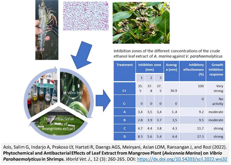
Phytochemical and Antibacterial Effects of Leaf Extract from Mangrove Plant (Avicennia Marina) on Vibrio Parahaemolyticus in Shrimps
Azis, Salim G, Indarjo A, Prakoso LY, Hartati R, Daengs AGS, Meiryani, Aslan LOM, Ransangan J, and Rozi.
World Vet. J. 12(3): 260-265, 2022; pii:S232245682200032-12
DOI: https://dx.doi.org/10.54203/scil.2022.wvj32
ABSTRACT: Recently, there has been a tremendous increase in the studies addressing the application of bioactive compounds from the natural ecosystem, particularly for medical purposes. Hence, the present study investigated the antibacterial properties of the secondary metabolites possibly contained in the leaves of Avicennia marina (A. marina) for possible prevention of Vibrio parahaemolyticus (V. parahaemolyticus), a devastating bacterial pathogen in shrimp aquaculture. In the current study, secondary metabolites were extracted from the leaves of mangrove plant using ethanol extraction method. The ethanolic extracts were then subjected to phytochemical and antibacterial activity tests. The results from the phytochemical analysis demonstrated that the ethanolic extract from the mangrove plant contained varying amounts of flavonoids, tannins, saponins, polyphenols, alkaloids, steroids, and triterpenoids. However, the number of flavonoids and alkaloids seemed to be higher than the other metabolites. The antibacterial activity analysis through the agar diffusion method has shown that different concentrations (50 ppm, 100 ppm, 200 ppm, and 300 ppm) of the ethanolic extract of A. marina inhibited the V. parahaemolyticus. At 300 ppm, the plant extract exhibited 17.3% antibacterial effectiveness, compared to the antibacterial activity of chloramphenicol. The findings indicated that the secondary metabolites of A. marina have the potential that can be developed as an alternative treatment for aquatic animal diseases in the future.
Keywords: Aquaculture, Bioactive compounds, Mangrove ecosystem, Treatment
[Full text-PDF] [Crossref Metadata] [Scopus] [Export from ePrint] [How to Cite]
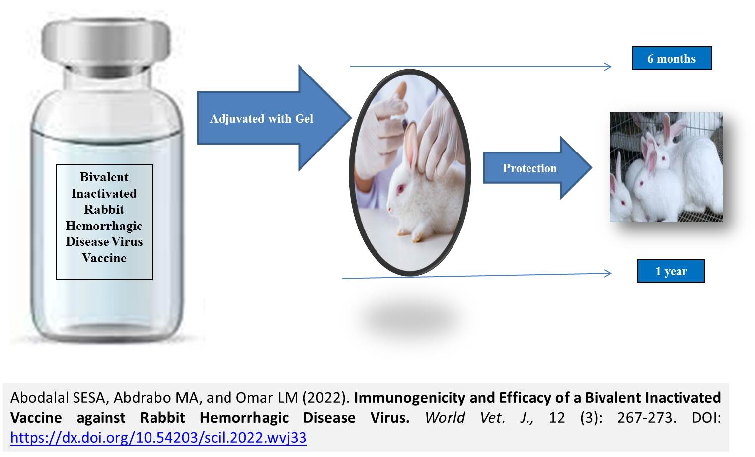
Immunogenicity and Efficacy of a Bivalent Inactivated Vaccine against Rabbit Hemorrhagic Disease Virus
Abodalal SEA, Abdrabo MA, and Omar LM.
World Vet. J. 12(3): 266-273, 2022; pii:S232245682200033-12
DOI: https://dx.doi.org/10.54203/scil.2022.wvj33
ABSTRACT: Rabbit hemorrhagic disease is a fatal threat to rabbits that causes sustainability problems and substantial economic losses. The aim of the current study was to compare the immuno-enhancing effects of a bivalent inactivated rabbit hemorrhagic disease virus (RHDV) vaccine adjuvanted with Montanide with an inactivated RHDV vaccine with an aluminum hydroxide gel. Montanide incomplete seppic adjuvant 71 VG was prepared as an oil emulsion, and several batches adjuvanted with an aluminum hydroxide gel were prepared. Then, 250 New Zealand rabbits aged 6 weeks were randomly allocated to three groups. Group 1 was subjected to the bivalent inactivated RHDV adjuvanted with an aluminum hydroxide gel, Group 2 received the oil-emulsion vaccine adjuvanted with Montanide, and Group 3 was left unvaccinated as a negative control group. Efficacy was determined using a hemagglutination inhibition test, and resistance was determined using virulent RHDVa and RHDV2. The clinical signs included sudden death, nervous manifestations, aimless running, lateral recumbence, and crying before death. The mortality rates were recorded at 3 weeks, 3 months, 6 months, and 12 months after vaccination. In addition, blood samples were collected on the first day as well as 1, 2, 3, 4, 6 weeks post-vaccination (WPV), and 2, 3, 4 month post-vaccination (MPV) until 12 MPV. Serological analysis indicated that the bivalent inactivated RHDV oil-emulsion vaccine was more effective than the bivalent inactivated RHDV aluminum hydroxide gel vaccine, resulting in improved immune responses and longer-lasting protective immunological responses in vaccinated rabbits. The bivalent inactivated RHDV oil-emulsion vaccine was also sterile and safe and helped the protection of the rabbits against RHDVa and RHDV2, hence reducing the time and effort required during the vaccination process and reducing the levels of discomfort for the rabbits.
Keywords: Immunity, Inactivated vaccine, Oil emulsion, Rabbit hemorrhagic disease virus
[Full text-PDF] [Crossref Metadata] [Scopus] [Export from ePrint] [How to Cite]
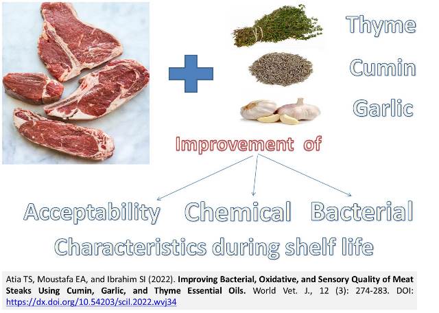
Improving Bacterial, Oxidative, and Sensory Quality of Meat Steaks Using Cumin, Garlic, and Thyme Essential Oils
Atia TS, Moustafa EA, and Ibrahim SI.
World Vet. J. 12(3): 274-283, 2022; pii:S232245682200034-12
DOI: https://dx.doi.org/10.54203/scil.2022.wvj34
ABSTRACT: The meat industry increasingly considers meat shelf life as a critical problem. Some essential oils contain antibacterial and antioxidant characteristics that help to keep meat safe. Therefore, the purpose of this study was to evaluate the preservation benefits, including antibacterial and antioxidant properties, of cumin, garlic, and thyme essential oils at 1% on chilled beef meat steaks, as well as their effects on pH, total volatile basic nitrogen (TVBN), thiobarbituric acid (TBA), and related sensory aspects (color, odor, appearance, consistency, and overall acceptability). The results of the current study showed that pretreating beef meat steaks with EOs of cumin, garlic, and thyme at a concentration of 1% effectively reduced levels of APC, coliform count, staph aureus count, TVBN, and TBA while extending shelf life to 12, 15, and 18 days when stored at 4°C. In terms of antibacterial and antioxidant properties, shelf life, and sensory quality on beef meat steaks, the thyme essential oil group outperformed cumin and garlic essential oils. The current study introduced an effective natural preservative alternative that could replace undesirable synthetic substances in the future while also lowering antibiotic resistance.
Keywords: Coliforms, Cumin, Garlic, Preservation, Shelf life, Thyme
[Full text-PDF] [Crossref Metadata] [Scopus] [Export from ePrint] [How to Cite]
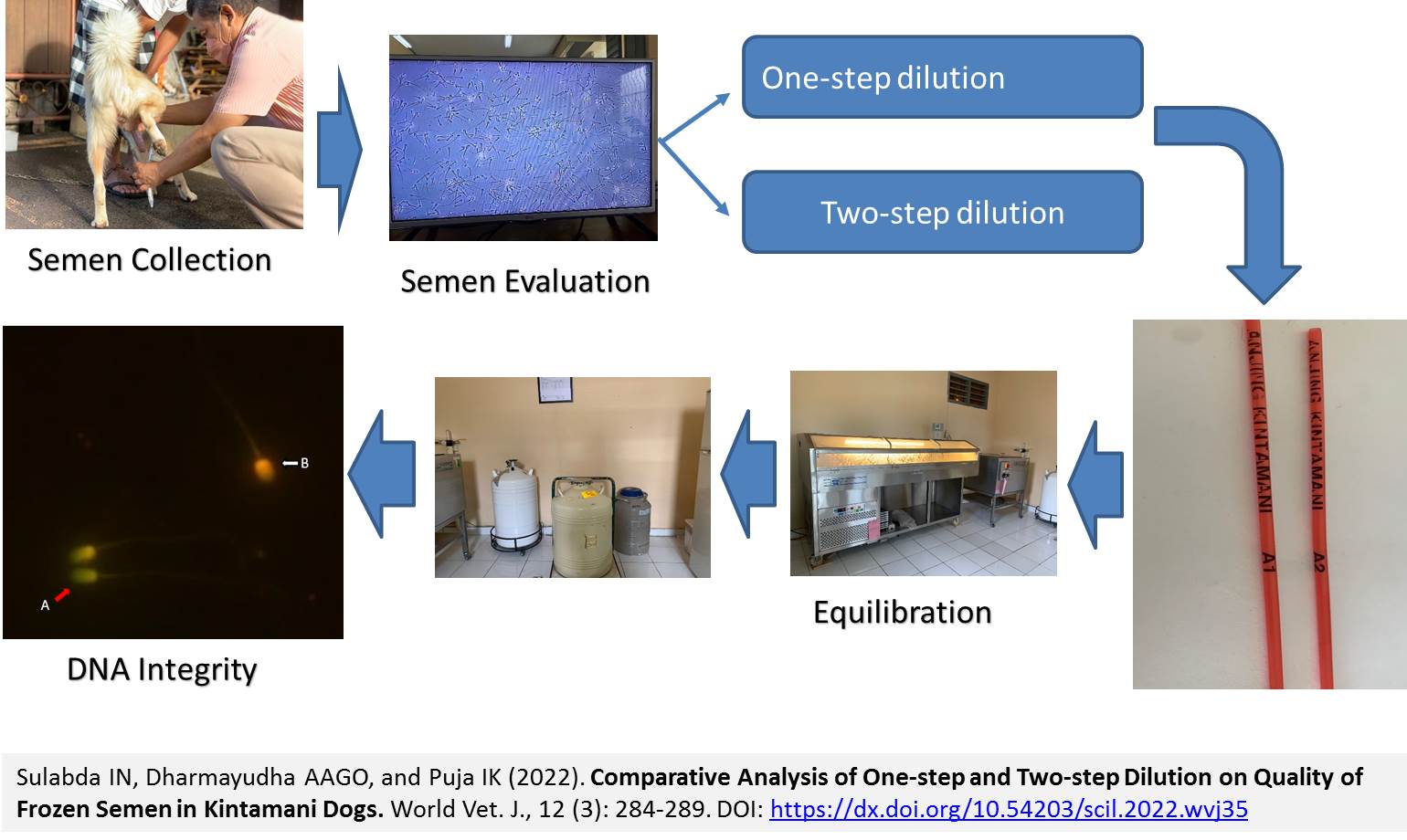
Comparative Analysis of One-step and Two-step Dilution on Quality of Frozen Semen in Kintamani Dogs
Sulabda IN, Dharmayudha AAGO, and Puja IK.
World Vet. J. 12(3): 284-289, 2022; pii:S232245682200035-12
DOI: https://dx.doi.org/10.54203/scil.2022.wvj35
ABSTRACT: Preservation of sperm by freezing allows breeding dogs that are separated over long distances. To increase the fertility of frozen and then thawed spermatozoa, they must be able to survive the process. The current study aimed to evaluate the sperm motility and DNA integrity of Kintamani dogs extended in extenders with one-step and two-step dilution techniques. Ejaculates collected from four dogs were used in the current study. The semen was divided into two equal parts and diluted with extenders using two different dilution techniques, namely One-step dilution in Tris egg yolk containing 7% glycerol, and a two-step dilution technique diluted in an initial 2:1 with an extender, containing 20% egg yolk without glycerol. The same volume of the second extender was added, including 14% glycerol. The sample was loaded into 0.25 ml straws, cooled to 4°C for 4 hours, equilibrated, and then plunged into the liquid nitrogen. The sperm motility was evaluated using Computer-Assisted Sperm Analysis (CASA), and DNA integrity was assessed using Acridine Orange (AO) stained. Results showed that the sperm motility of Kintamani dogs in extenders using two-step dilution was significantly higher compared to the one-step dilution technique. In addition, the obtained results indicated that two types of dilution steps in Kintamani dog semen were not detrimental to the sperm DNA integrity during the freezing process. In conclusion, extenders with two types of dilution techniques could maintain sperm motility above 30%, and no difference between one and two steps dilution was detected.
Keywords: Dilution techniques, DNA integrity, Egg yolk, Kintamani dog, Motility
[Full text-PDF] [Crossref Metadata] [Scopus] [Export from ePrint] [How to Cite]
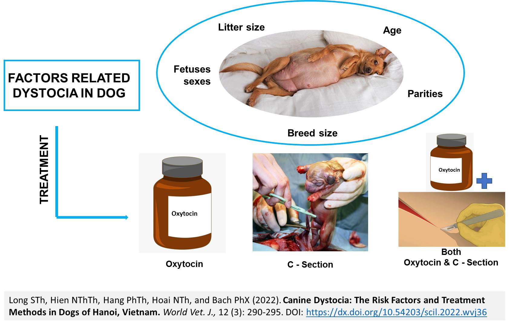
Canine Dystocia: The Risk Factors and Treatment Methods in Dogs of Hanoi, Vietnam
Long STh, Hien NThTh, Hang PhTh, Hoai NTh, and Bach PhX.
World Vet. J. 12(3): 290-295, 2022; pii:S232245682200036-12
DOI: https://dx.doi.org/10.54203/scil.2022.wvj36
ABSTRACT: Dystocia is a common disorder that can cause harmful health risks to bitch and puppies. The aim of the current study was to evaluate some risk factors related to canine dystocia and the application of treatment methods to 612 diagnosed cases in Gaia Pets Clinic and Resort, Hanoi, Vietnam, from December 2013 to May 2020. The investigated factors comprised age, parity and breed size, and litter size, as well as fetal sex in relation to the proportion of dystocia in female canines. Dystocia was frequently observed in female dogs aged 1-3 years, with rates of 76.1%. The highest proportion of dystocia was found in the first litter group (80.21%). The incidence of dystocia increased as the weight of the dog decreased, and it was prevalent in the small breed (61.93%). Dystocia risk decreased as the litter size increased. The interventions used in this study were medical treatment with the hormone oxytocin (1.8%), surgical management with cesarean section (86.11%), and a combination of oxytocin and cesarean section (12.09%), with the success rates of each treatment method as 100%, 98.86%, and 100%, respectively. Some risk factors, such as age, parity, breed size, and litter size identified in the present research, could be used as prognostic indicators in the veterinary practice to optimize the survival rate of female dogs and puppies.
Keywords: Age, Breed, Dystocia, Fetus sex, Litter size, Parities
[Full text-PDF] [Crossref Metadata] [Scopus] [Export from ePrint] [How to Cite]
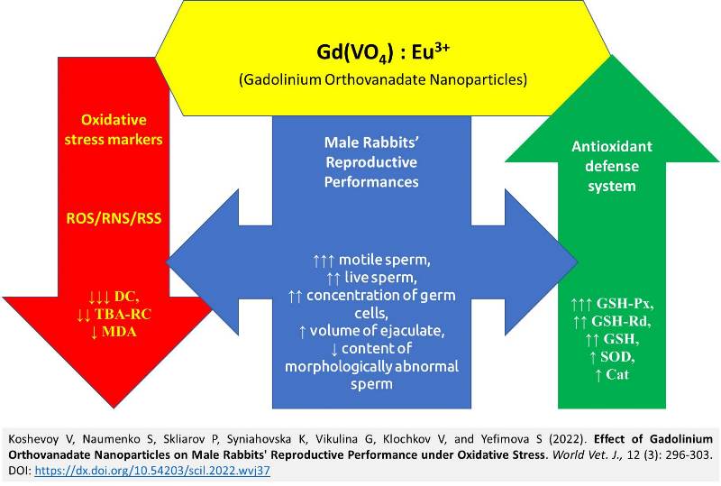
Effect of Gadolinium Orthovanadate Nanoparticles on Male Rabbits' Reproductive Performance under Oxidative Stress
Koshevoy V, Naumenko S, Skliarov P, Syniahovska K, Vikulina G, Klochkov V, and Yefimova S.
World Vet. J. 12(3): 296-303, 2022; pii:S232245682200037-12
DOI: https://dx.doi.org/10.54203/scil.2022.wvj37
ABSTRACT: Oxidative stress as a leading factor of male infertility requires correction with modern pharmacological agents, particularly redox-active nanoparticles, to improve sperm quality and hormonal balance. The current experimental study aimed to investigate the effect of orthovanadate nanoparticles of rare earth elements, particularly Gadolinium, with pronounced redox properties on the reproductive function of male rabbits under oxidative stress. A total of 36 mature male Hyla rabbits were divided into three groups of intact control (n = 12) and two experimental groups, including rabbits ubder oxidative stress (n = 12), induced by the introduction of tert-Butyl hydroperoxide, and those under oxidative stress plus hydrosol of gadolinium orthovanadate nanoparticles (NPs, n = 12) intake for 14 days. There were four rabbits per three replicates in each group. Animals of all groups were kept on the same diet and had free access to water. The use of NPs led to an improvement in sperm quality indicators. There was an improvement in motility and ejaculate volume indicators (by 14.6% and 39.2%, respectively), a reduction of the content of morphologically abnormal sperm by 26.7%; normalization of sex hormones, an increase in the level of total testosterone (by 113%) with a decrease in 17-β-estradiol (by 16.5%). This sex hormones improvement led to an increase in the androgen saturation of the rabbit’s body (free androgen index at the end of the experiment was 36.5%). The obtained changes were accompanied by a decrease in the oxidative load, as evidenced by a reduced content of diene conjugates and thio-barbituric acid-reactive compounds in the blood serum of rabbits by 30.4% and 26.8%, compared to the control. At the same time, there was an increase in the antioxidant potential, especially its glutathione link – the activity of glutathione peroxidase and glutathione reductase (by 42.5% and 34.2%, respectively), and the content of reduced glutathione increased by 62.3%, compared to the indicators before the introduction of NPs. The results of the study confirmed the effectiveness of using gadolinium orthovanadate NPs to correct the reproductive function of males under oxidative stress.
Keywords: Gadolinium orthovanadate, Male rabbits, Nanoparticles, Oxidative stress, Reproductive performances
[Full text-PDF] [Crossref Metadata] [Scopus] [Export from ePrint] [How to Cite]
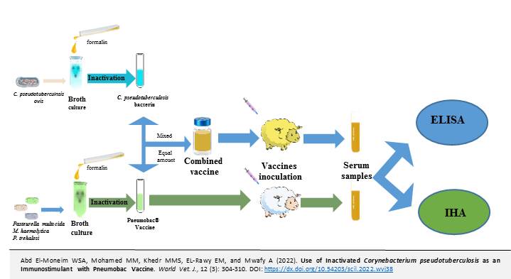
Use of Inactivated Corynebacterium pseudotuberculosis as an Immunostimulant with Pneumobac Vaccine
Abd El-Moneim WSA, Mohamed MM, Khedr MMS, EL-Rawy EM, and Mwafy A.
World Vet. J. 12(3): 304-310, 2022; pii:S232245682200038-12
DOI: https://dx.doi.org/10.54203/scil.2022.wvj38
ABSTRACT: Sheep breeders in Egypt suffer from pneumonic pasteurellosis caused by Pasteurella trehalosi, Pasteurella multocida, and Mannheimia haemolytica. The disease is responsible for significant economic losses in the sheep industry according to the high mortality rate and reduced carcass values. Pneumobac® is the primary vaccine in Egypt used to control pasteurellosis in sheep. Therefore, the aim of the present study was to estimate the nonspecific immune stimulating impact of Corynebacterium pseudotuberculosis ovis against Pasteurella in sheep vaccinated with Pneumobac®. Nine sheep were classified into three groups, each with three animals. The sheep in the first and second groups were inoculated with the inactivated culture of Pneumobac® and a combined inactivated culture of Pneumobac® with Corynebacterium pseudotuberculosis ovis bacterin, respectively. The third group was nonvaccinated and kept in control. Indirect haemagglutination test (IHA) and enzyme-linked immunosorbent assay (ELISA) were used to measure the humoral immune response to the produced vaccines. The results of the present study confirmed that the antibodies titer against Pasteurella multocida type A, D, and B6, Pasteurella trehalosi type T, and Mannheimia haemolytica type A significantly increased in sheep vaccinated with a combined vaccine (Pneumobac® and Corynebacterium pseudotuberculosis ovis bacterin), compared to those vaccinated with Pneumobac® alone. It was concluded that the addition of Corynebacterium pseudotuberculosis ovis bacterin to inactivated Pneumobac® vaccine could increase the immune response against pneumonic pasteurellosis.
Keywords: Corynebacterium pseudotuberculosis, Pasteurella multocida, Pasteurellosis, Pneumobac®
[Full text-PDF] [Crossref Metadata] [Scopus] [Export from ePrint] [How to Cite]
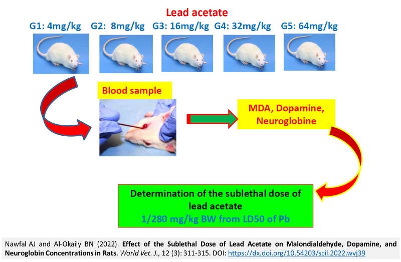
Effect of the Sublethal Dose of Lead Acetate on Malondialdehyde, Dopamine, and Neuroglobin Concentrations in Rats
Nawfal AJ and Al-Okaily BN.
World Vet. J. 12(3): 311-315, 2022; pii:S232245682200039-12
DOI: https://dx.doi.org/10.54203/scil.2022.wvj39
ABSTRACT: Lead can have detrimental behavioral, biochemical, and physiological effects on the body. The current experiment was designed to estimate the sublethal dose of lead acetate that induce oxidative stress on the central nervous system (CNS) in adult using the probit analysis. Moreover, the current study examined the dose-response curve by successive doses of lead acetate on some parameters related to oxidative stress for 28 days. A total of 36 adult male rats were randomly selected and divided equally into six experimental groups and treated for 28 days. Rats in the control group received distilled sterile water, and those in G1, G2, G3, G4, and G5 were gavaged with 4, 8, 16, 32, and 64 mg/kg of lead acetate, respectively. The result indicated a positive correlation between the successive doses of lead acetate. Malondialdehyde concentration decreased dopamine and neuroglobin by increasing the dose of lead acetate in experimental groups (G3, G4, and G5), compared to the control group. In conclusion, exposure to the sublethal dose of 16 mg/kg of lead acetate significantly alters the levels of the neurotransmitters and increases the production of oxidative stress in the CNS tissue.
Keywords: Central nervous system, Dopamine and Neuroglobin, Lead acetate, Malondialdehyde, Rat
[Full text-PDF] [Crossref Metadata] [Scopus] [Export from ePrint] [How to Cite]
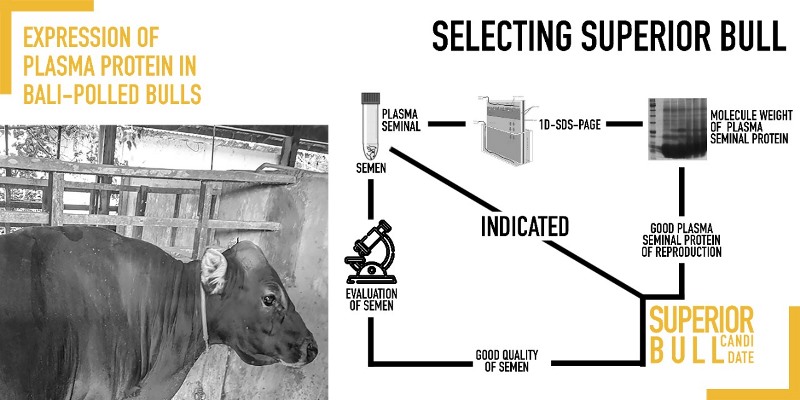
The Expression of Plasma Protein in Bali-polled Bulls Using 1D-SDS-PAGE
Diansyah AM, Yusuf M, Toleng AL, Dagong MIA, and Maulana T.
World Vet. J. 12(3): 316-322, 2022; pii:S232245682200040-12
DOI: https://dx.doi.org/10.54203/scil.2022.wvj40
ABSTRACT: The fertility rate of bulls in a breeding program is not only described by the quantity and quality of semen. Factors, such as the interstice factor of the sperm and the plasma component of semen, affect the fertility rate of bulls. The fertility rate can also be determined by identifying the protein content of semen plasma. Therefore, the current study aimed to identify the relationship between seminal plasma protein molecular weight and semen quality of Bali-polled bulls. The study was conducted at the Laboratory of Semen Processing, Faculty of Animal Science, Hasanuddin University, Makassar, Indonesia, the Research Center for Applied Zoology, National Research and Innovation Agency, Cibinong, Indonesia and the Laboratory of Animal Biotechnology Center, IPB University, Bogor, Indonesia from November 2021 to January 2022. The samples came from 5 Bali-polled and 5 Bali-horned bulls. Semen collection was conducted twice a week using an artificial vagina. The concentration of seminal plasma protein was determined by the Bradford method of 1D-SDS-PAGE. The study results showed that fresh semen of Bali-polled and Bali-horned bulls was considered a normal category. Seminal plasma proteins of Bali-polled and Bali-horned bulls were classified using 8 bands to categorize molecular weight; 150 kD (IGF-1), 110 kD (A-kinase anchoring protein 3), 93 kD (A-kinase anchoring protein 4), 54-87 kD (Arylsulfatase-a), 44-62 kD (N-Acetyl-ß-Guicosaminidase), 44kD (Phosphoglycerate kinase), 15-30 kD (BSP A1/A2, BSP-A3 and BSP-30 [BSP1, BSP3, and BSP5]) and 12-14 kD (Acidic seminal fluid proteins). The findings indicated that both Bali-polled and Bali-horned bulls could have a high reproductive rate. In conclusion, protein analysis based on molecular weight using 1D-SDS-PAGE can be used as a biomarker for semen quality in Bali-polled bulls. Therefore, evaluating the semen quality with a molecular basis as an additional indicator of superior bull in the selection process is an alternative method.
Keywords: Bali-polled bull, Seminal protein plasma, Sperm, 1D-SDS-PAGE
[Full text-PDF] [Crossref Metadata] [Scopus] [Export from ePrint] [How to Cite]
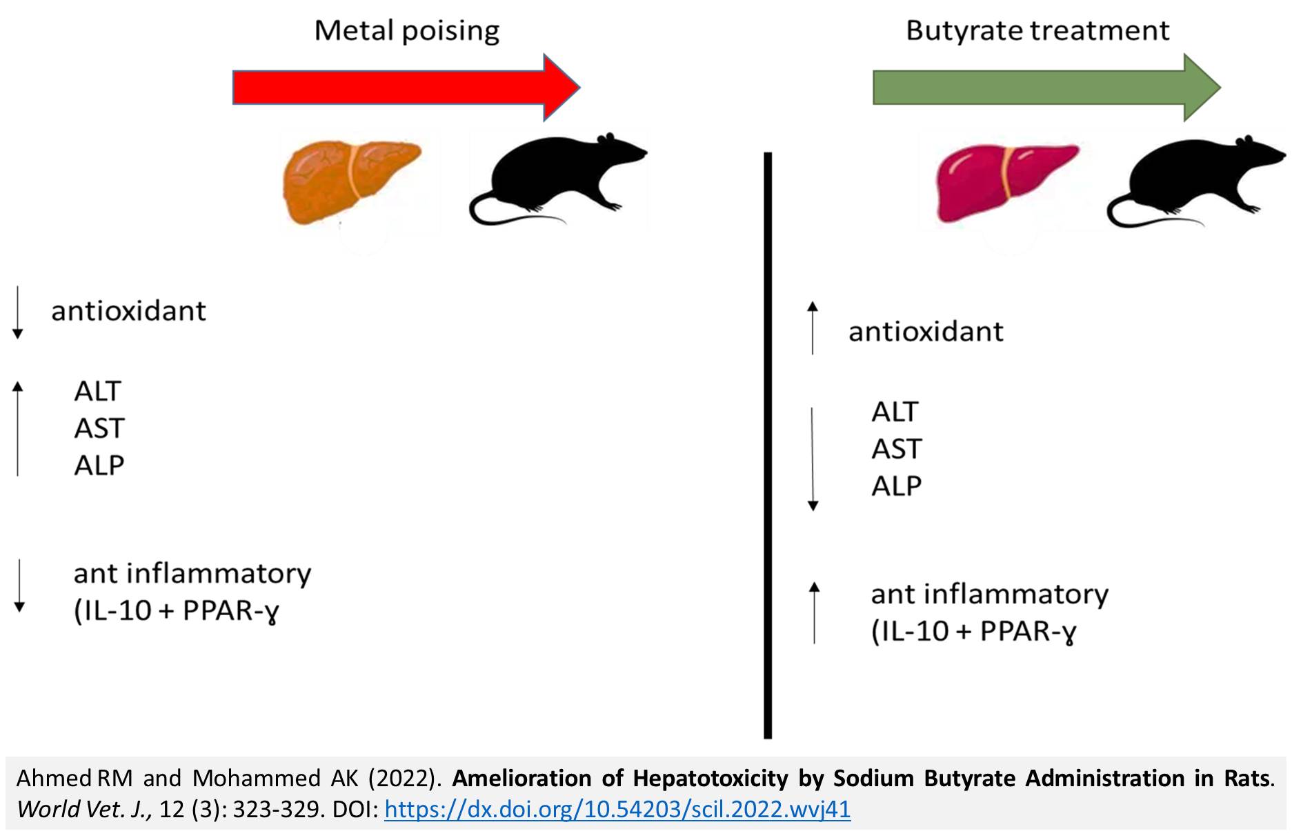
Amelioration of Hepatotoxicity by Sodium Butyrate Administration in Rats
Ahmed RM and Mohammed AK.
World Vet. J. 12(3): 323-329, 2022; pii:S232245682200041-12
DOI:https://dx.doi.org/10.54203/scil.2022.wvj41
ABSTRACT: Lead poisoning is a serious environmental issue with life-threatening consequences. Lead poisoning increases the risk of cancers, gastrointestinal disorders, hepatotoxicity, central nervous system diseases, nephropathy, and cardiovascular diseases in animals and humans. The current study aimed to investigate the effect of sodium butyrate, as an antioxidant, on protecting female adult rats from the harmful effects of lead acetate. A total of 40 adult female albino rats were divided randomly into four equal groups. The first group dealt as the control. The second group received lead acetate at a dose of 200 mg/kg daily orally. The third group received lead acetate at a dose of 50 mg/kg daily orally, and the fourth group received both sodium butyrate and lead acetate orally/day for 35 days. The result indicated that sodium butyrate reduced the concentration of liver enzymes (ALT, AST, and ALP) which were elevated by lead acetate poising. Moreover, sodium butyrate ameliorates the redux status by decreasing malondialdehyde and increasing total antioxidant capacity. Additionally, sodium butyrate-treated rats showed significant alterations in the expression of peroxisome proliferator-activated receptor gamma and interleukin -10 genes. In conclusion, this study reveals an unrecognized role for peroxisome proliferator-activated receptor gamma and Interleukin-10 signaling after sodium butyrate treatment in regulating the immunopathology that occurs during lead acetate poising.
Keywords: Interleukin-10, Lead acetate toxicity, Sodium butyrate, PPAR-gamma, Rat
[Full text-PDF] [Crossref Metadata] [Scopus] [Export from ePrint] [How to Cite]
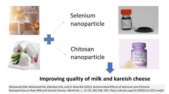
Antimicrobial Effects of Selenium and Chitosan Nanoparticles on Raw Milk and Kareish Cheese
Mohamed ShN, Mohamed HA, Elbarbary HA, and EL-Roos NA.
World Vet. J. 12(3): 330-338, 2022; pii:S232245682200042-12
DOI: https://dx.doi.org/10.54203/scil.2022.wvj42
ABSTRACT: The contamination of milk and its dairy products with different microorganisms could cause public health hazards. Antibacterial nanoparticles (NPs) are a novel way to ensure that milk and milk products are safe. The present study investigated the effect of chitosan NPs (CS-NPs) and selenium NPs (Se-NPs) on some microorganisms, which consequently affect raw milk and Kareish cheese. Small-sized nanomaterials of Se-NPs and CS-NPs at the size of approximately 20 nm were used in this study. The samples were 700 ml raw milk and 700g Kareish cheese manufactured from 3000 mg milk. The concentrations of used nanoparticles were 0.5%, 1%, and 1.5% for Se-NPs and 2.5%, 5%, and 10% for CS-NPs. They were used to improve the microbial properties of milk and Kareish cheese samples during storage at the refrigerated temperature of 4°C. The aerobic plate count, Enterobacteriaceae count, Staphylococcus count, and mold count were significantly reduced in milk and Kareish cheese samples treated with CS-NPs and Se-NPs. The study has confirmed that CS-NPs and Se-NPs indicated high antimicrobial activity against the studied microorganisms at all concentrations although CS-NPs were more effective than Se-NPs. It can be concluded that these NPs can be used as preservatives in milk and milk products, such as Kareish cheese. In addition, increasing the concentrations of these NPs by 10% for CS-NPS and 1.5% for Se-NPS boosted their effects.
Keywords: Chitosan, Enterobacteriaceae, Kareish cheese, Nanoparticle, Selenium, Staphylococcus aureus
[Full text-PDF] [Crossref Metadata] [Scopus] [Export from ePrint] [How to Cite]
Basic Principles and Applications of Live Cell Microscopy Techniques: A Review
Mekuria TA and Kinde MZ.
World Vet. J. 12(3): 339-346, 2022; pii:S232245682200043-12
DOI: https://dx.doi.org/10.54203/scil.2022.wvj43
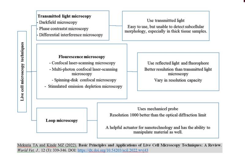
ABSTRACT: Live cell imaging has provided great benefits in studying multiple processes and molecular interactions within and/or between cells. This review aimed to describe the common live cell microscopy techniques and briefly explain their principles and applications. A wide range of microscopic techniques, from conventional transmitted light to an array of fluorescence microscopy techniques, including advanced super-resolution techniques, can be applied for live-cell imaging. Transmitted light microscopy uses focused transmitted light that goes through a condenser to achieve a very high illumination on the specimen. On the other hand, fluorescence microscopy uses reflected light to capture images of cells or molecules that have been fluorescently dyed. Techniques for transmitted light microscopy are simple to use but have poor resolution. Although the resolution of fluorescent microscopy techniques is only approximately 200-300 nm, this is nevertheless an improvement over conventional transmitted methods. Conventional light microscopy’s resolution was improved by the introduction of the super-resolution microscopy technology family. These methods “break” the diffraction limit, enabling fluorescence imaging with resolutions up to ten times higher than those possible with traditional methods. Each live cell imaging method has advantages and drawbacks. The primary deciding criteria for choosing the type of microscope are the study’s objectives, previous experience, the researcher’s interests, and financial viability. Hence, a thorough understanding of the technique and application of the various live-cell microscopy methods is paramount in life science studies.
Keywords: Application, Fluorescence, Imaging, Laser-Scanning, Live cell, Microscopy
[Full text-PDF] [Crossref Metadata] [Scopus] [Export from ePrint] [How to Cite]
Previous issue | Next issue | Archive
This work is licensed under a Creative Commons Attribution 4.0 International License (CC BY 4.0).


