 |
|  |
| 
Previous issue | Next issue | Archive
![]() Volume 12 (4); December 25, 2022 [Booklet] [EndNote XML for Agris]
Volume 12 (4); December 25, 2022 [Booklet] [EndNote XML for Agris]
 Research Paper
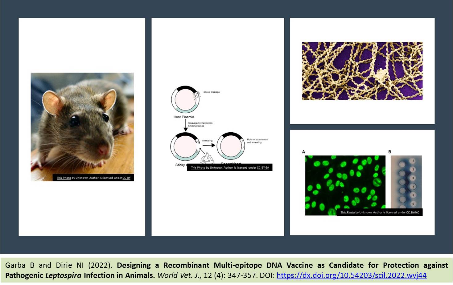
 Designing a Recombinant Multi-epitope DNA Vaccine as Candidate for Protection against Pathogenic Leptospira Infection in Animals
Garba B and Dirie NI.
World Vet. J. 12(4): 347-357, 2022; pii:S232245682200044-12
DOI: https://dx.doi.org/10.54203/scil.2022.wvj44
ABSTRACT: Leptospirosis can cause severe disease and probable death in humans. Antigenic epitopes from pathogenic strains of the bacteria have shown potential for serving as vaccine candidates and play a key role in the sensitivity and specificity of immunodiagnostic tests. This in-vitro analysis was undertaken to develop a prototype recombinant DNA vaccine using in-silico epitope prediction method. Epitope prediction software programs predicted the most antigenic linear B-cell epitopes of OmpL1, LipL32, LipL41, Loa22, and LigA. Thirteen epitopes were predicted, connected by the Gly-Ser linker, and synthesized. The purity of the concentrated recombinant multi-epitope protein was assessed by restriction enzyme digestion and gel electrophoresis. In-vitro expression on mammalian Chinese Hamster Ovary cell line indicated strong cytoplasmic fluorescence produced based on an indirect immunofluorescence antibody test. The green color of the cytoplasm indicates successful transcribed and translated DNA as against the blue-stained nucleus observed in the un-transfected control group based on the indirect immunofluorescence antibody test. The findings of the current study showed high antibody binding potentials of the vaccine constructs, which could be used for diagnostic applications or as polyvalent vaccine candidates.
Keywords: B-cell epitopes, Indirect immunofluorescence antibody test, Leptospirosis, Multi-epitope vaccine, Recombinant vaccine
[Full text-PDF] [Crossref Metadata] [Scopus] [Export from ePrint] [How to Cite]
 Research Paper
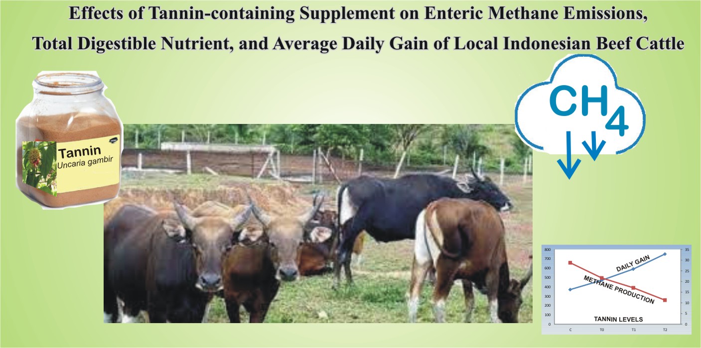
Effects of Tannin-containing Supplement on Enteric Methane Emissions, Total Digestible Nutrient, and Average Daily Gain of Local Indonesian Beef Cattle
Ramaiyulis R, Metri Y, Irda I, Kurnia D, and Syukriani D.
World Vet. J. 12(4): 358-362, 2022; pii:S232245682200045-12
DOI: https://dx.doi.org/10.54203/scil.2022.wvj45
ABSTRACT: Reducing methane (CH4) emissions is one of the most critical goals in ruminant nutrition. This study aimed to evaluate the effect of concentrate and tannin supplementation on the mitigation of methane gas in Indonesian local beef cattle. The current study was conducted in vivo using 12 Bali cattle using a completely randomized design with four treatments and three replicates. Cattle were fed a basal ration with field grass (control), the addition of concentrate 25% dry matter (DM) ration no tannin as well as tannin supplemented in concentrate at levels of 0.12% and 0.18% of DM concentrate. The concentrate contains 7.5% crude protein and 71.25% total digestible nutrients and tannin supplementation using gambir (Uncaria gambir Indonesia) tannin extract. The parameters measured were apparent digestibility, total digestible nutrients, methane production, and average daily gain. The results showed that concentrate addition significantly increased DM consumption, crude protein digestibility, and total digestible nutrients. Supplementation of 0.18% tannin in concentrate can mitigate 49.7% methane gas production resulting in energy efficiency, which was reflected in the weight gain rate of 0.75 kg/day. In conclusion, present results suggest that the supplementation of 0.18% gambier tannin extract in concentrate could be considered a suitable feed additive to mitigate methane gas production and increase the average daily gain of Indonesian local beef cattle.
Keywords: Digestibility, Feed supplement, Gambir, Methane, Tannin
[Full text-PDF] [Crossref Metadata] [Scopus] [Export from ePrint] [How to Cite]
 Research Paper
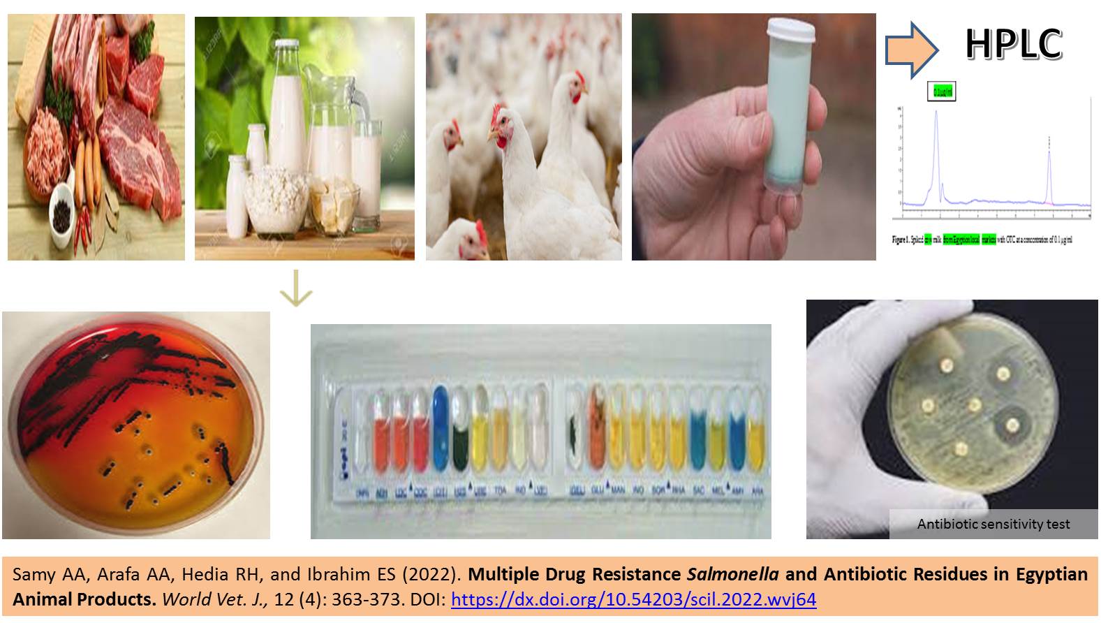
Multiple Drug Resistance Salmonella and Antibiotic Residues in Egyptian Animal Products
Samy AA, Arafa AA, Hedia RH, and Ibrahim ES.
World Vet. J. 12(4): 363-373, 2022; pii:S232245682200064-12
DOI: https://dx.doi.org/10.54203/scil.2022.wvj64
ABSTRACT: Food of animal origin is considered a primary source of foodborne diseases. The misuse of antibiotics to treat and control many bacterial diseases in farm animals has led to multiple antibiotic-resistant pathogens in contaminated food that can seriously threaten public health. The present study aimed to highlight the impact of antimicrobial misuse in Egyptian beef meat, poultry, and dairy farms on the emergence of multiple antibiotic resistance Salmonella and the detection of antibiotic residues in milk. A total of 1050 samples were collected randomly from poultry (liver, intestinal content, and bone marrow), meat, and milk products from different Egyptian governorates. Salmonellae were isolated from the collected samples and subjected to antimicrobial sensitivity testing through disk diffusion test using the most commonly used seven antibiotics in veterinary fields (cefradine, ciprofloxacin, oxytetracycline, erythromycin, amoxicillin, ampicillin, and streptomycin). The detection of oxytetracycline residue in milk samples was performed by high-performance liquid chromatography (HPLC). Most isolated Salmonellae were multiple drug resistant with an incidence rate of 8.6%, 15.4%, and 4% from poultry, meat-associated products, and milk-associated products, respectively, from different governorates. Antibiogram test showed that the isolated Salmonella from poultry, meat, and milk samples were resistant to oxytetracycline at 100%, 31.4%, and 43%, to amoxicillin at 73.3%, 31%, and 50%, and to ampicillin 66.6%, 50%, and 57%, respectively. No resistance to ciprofloxacin was detected in Salmonella isolates from all samples. Using HPLC, oxytetracycline residues were detected in milk samples. In conclusion, more attention should be paid to the connection between the widespread emergence of antibiotic-resistant Salmonella in Egypt and the overuse of antimicrobials in poultry, dairy, and meat farms. This connection affects consumer health and increases the likelihood of resistance genes spreading between different bacterial species.
Keywords: Antibiogram, High-performance liquid chromatography, Multiple drug resistance, Salmonella
[Full text-PDF] [Crossref Metadata] [Scopus] [Export from ePrint] [How to Cite]
 Research Paper
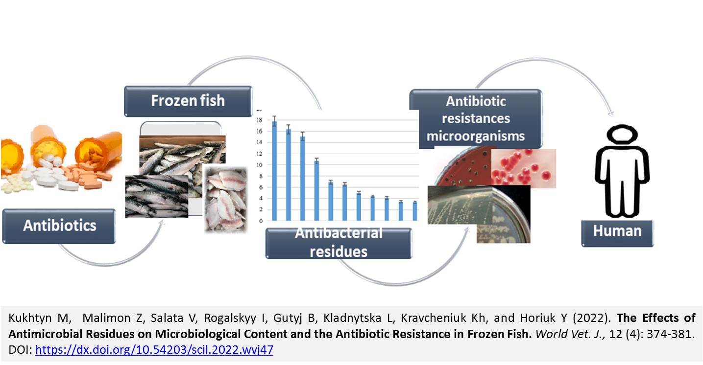
The Effects of Antimicrobial Residues on Microbiological Content and the Antibiotic Resistance in Frozen Fish
Kukhtyn M, Malimon Z, Salata V, Rogalskyy I, Gutyj B, Kladnytska L, Kravcheniuk Kh, and Horiuk Y.
World Vet. J. 12(4): 374-381, 2022; pii:S232245682200047-12
DOI: https://dx.doi.org/10.54203/scil.2022.wvj47
ABSTRACT: As fish are perishable foods, their storage conditions require appropriate sanitary and temperature regimes. The producers commonly use various antibiotics to stop fish’s microbiological and biochemical processes. The current research aimed to examine antibacterial residues in frozen fish (Argentina, flounder, lackerda, mackerel, capelin, salka, saithe, herring, dorado, and pink salmon) to find their influence on the quantitative content of microorganisms and to determine the sensitivity of isolated psychrotrophic bacteria to antibiotics. A total of 75 samples were collected from the fillets of frozen fish species. These fish were imported from Norway (16 samples), Vietnam (24 samples), Russian Federation (8 samples), China (14 samples), New Zealand (2 samples), Italy (2 samples), United States (4 samples), and United Kingdom (5 samples). The obtained results revealed that aminoglycosides (Gentamicin, Kanamycin, Spectinomycin, Dihydrostreptomycin, Paromomycin, and Apramycin) were in 45.6 ± 1.4% of frozen fish. The findings indicated the presence of some antibacterial residues (Nalidixic acid, antibiotics: Apramycin, Kanamycin, Tiamulin, and Nafcillin) in frozen fish, the definition of which has not been specified in the EU Regulation. This gives grounds to prohibit the use or develop standards for the maximum permissible concentration of these antibacterial substances in fish. The most common psychrotrophic bacteria isolated from frozen fish without antibacterial residues were highly sensitive to antibiotics, including Penicillin, Tetracycline groups, and Aminoglycosides. Therefore, it can be concluded that the residual levels of various biocides found in fish are a source for the expression of multi-resistance genes, which can be transmitted to consumers in the food chain.
Keywords: Antibacterial residues, Antibiotic resistances, Frozen fish, Multi-resistance genes, Psychrotrophic microorganisms
[Full text-PDF] [Crossref Metadata] [Scopus] [Export from ePrint] [How to Cite]
 Research Paper
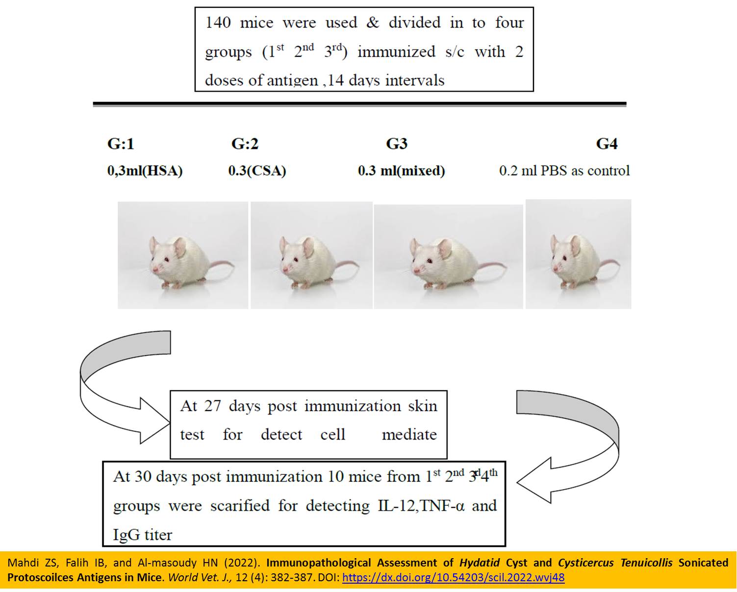
Immunopathological Assessment of Hydatid Cyst and Cysticercus Tenuicollis Sonicated Protoscoilces Antigens in Mice
Mahdi ZS, Falih IB, and Al-masoudy HN.
World Vet. J. 12(4): 382-387, 2022; pii:S232245682200048-12
DOI: https://dx.doi.org/10.54203/scil.2022.wvj48
ABSTRACT: The present study was designed to investigate the cross-protection (protective immunity) between Hydatid cyst and Bladder worm and evaluate the immunologic response of both humerol and cellular immunity in mice. To achieve these goals, 120 mice were used and equally divided into four groups immunized subcutaneously with 2 doses of antigen at the first and 14 days of the experiment. Mice in the first group (n=30) were immunized with 0.3 ml of hydatid cyst sonicated protoscolex antigen. Those in the second group (n=30) were immunized s/c with 0.3 ml of Cysticercus tenuicollis sonicated protscolex antigen. The third group (n=30) was immunized with 0.3 ml of both antigens (0.15 + 0.15), and the fourth group was a control group in which the mice were intraperitoneally injected with 0.2 ml of phosphate buffer solution. At the end of the experiment (30 days), blood samples were taken from the hearts of mice in all groups after being anesthetized by intramuscular injection of Ketamine 60 mg/kg, and Xylazine 12 mg/kg for the assessment of mouse Interleukin-12, IgG, and tumor necrosis alpha levels. The skin test results 24 hours (day 28) post-immunization showed an increase in the skin thickness against both antigens in the treatments, compared to the control. However, there was a decrease at 48 hours (day 29) post-examination in all groups. The results of TNFα titer showed higher titer in the third group, compared to the first, second, and fourth groups. Interleukin 12 concentration showed a higher titer in the third group than in the first, second, and fourth groups. The IgG concentration showed higher titer in the third group compared to the first, second, and fourth groups. In conclusion, immunopathological studies have shown that Ags used in the study, induce humoral and cellular immunity, compared to each Ag alone, and the mixed antigens were much more immunogenic. This cross-reactivity and synergistic interactions between the two parasites may be the cause of their antigenic activities.
Keywords: Immunopathological cross reaction, Interlukin-12, Parasitic antigens, Sonicated protoscolex antigens, TNFα
[Full text-PDF] [Crossref Metadata] [Scopus] [Export from ePrint] [How to Cite]
 Research Paper
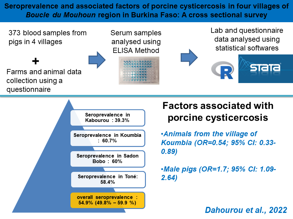
Seroprevalence and Associated Risk Factors of Porcine Cysticercosis in Boucle Du Mouhoun Region of Burkina Faso: A Cross-sectional Survey
Dahourou LD, Gbati OB, Nda KM, Tapsoba ASR, Traore A, Millogo A.
World Vet. J. 12(4): 388-394, 2022; pii:S232245682200049-12
DOI: https://dx.doi.org/10.54203/scil.2022.wvj49
ABSTRACT: Taenia solium cysticercosis is a neglected tropical zoonosis with economic and public health importance. Cysticercosis is widely present in low-income countries with extensive pig breeding systems and poor human hygiene practices. In Burkina Faso, a study on porcine cysticercosis has been done only in Boulkiemde province. There is a lack of serological data on this disease in other areas, such as Balés province in the Boucle du Mouhoun region. This study aimed to assess the prevalence and risk factors of porcine cysticercosis in Boucle du Mouhoun, Burkina Faso. A total of 373 serum samples were collected from local breed pigs that were randomly selected from four villages in the mentioned region. Data were also collected using a structured questionnaire to determine explanatory factors for the infection. Serum samples were tested using an antigen ELISA test to detect circulating antigens of Tænia solium. The prevalence of the disease was 54.9% (95% CI = 49.8-59.9). Following univariate and multivariate logistic regression analysis, pigs originating from Kombia were found to be a protective factor (OR=0.54, 95% CI = 0.33-0.89). Male pigs were more likely to be infected than females (OR= 1.7, 95% CI = 1.09-2.64). The prevalence and factors associated with Taenia solium cysticercosis were identified and according to these data, porcine cysticercosis had a high prevalence in this area. Therefore, it is important to implement control actions focusing on disease control and public health for people infected with Taenia solium.
Keywords: Burkina Faso, Prevalence, Pigs, Taenia solium, Zoonosis
[Full text-PDF] [Crossref Metadata] [Scopus] [Export from ePrint] [How to Cite]
 Research Paper
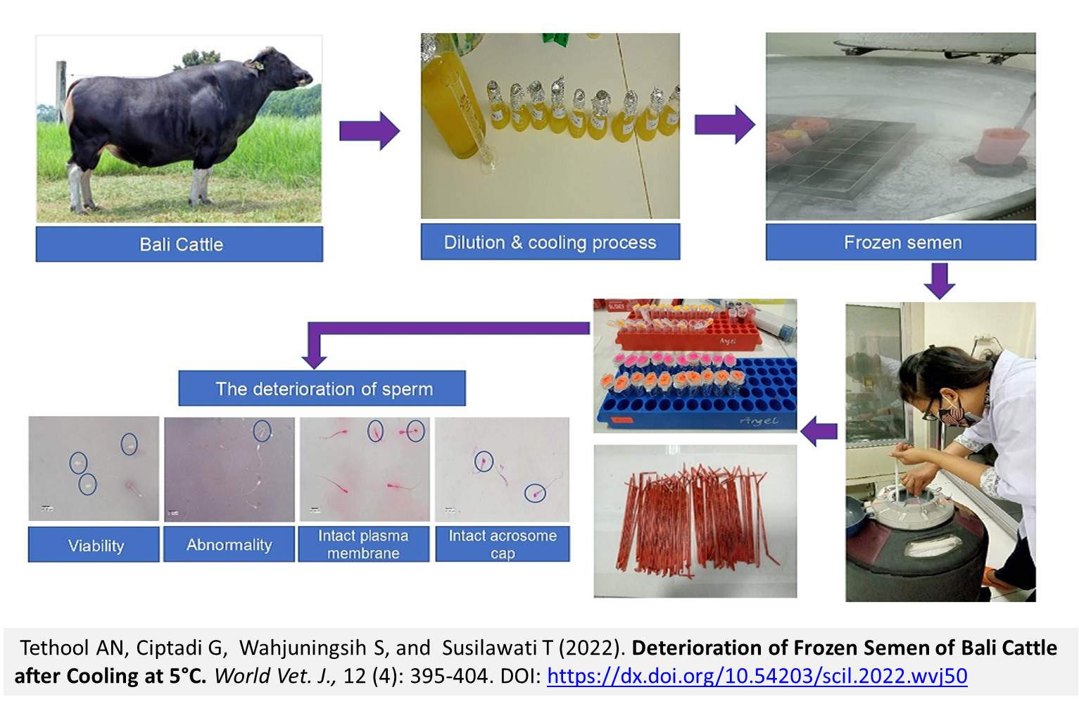
Deterioration of Frozen Semen of Bali Cattle after Cooling at 5°C
Tethool AN, Ciptadi G, Wahjuningsih S, and Susilawati T.
World Vet. J. 12(4): 395-404, 2022; pii:S232245682200050-12
DOI: https://dx.doi.org/10.54203/scil.2022.wvj50
ABSTRACT: Frozen semen is produced through several stages, which deteriorate spermatozoa. This research aimed to evaluate the deterioration degree of frozen semen after 5 °C cooling and freezing of Bali cattle. The samples included 10 male Bali cattle with a body weight of 542-668 kg, from which semen was collected once a week for five weeks. The deterioration of each individual’s sperm was determined by observing two distinct straws. The parameters observed included viability, abnormalities, intact plasma membrane, and intact acrosome cap. Initial observations of the parameters were conducted following the addition of semen to diluent A1 (AD) as much as the volume of fresh semen. The semen in the AD group was not cooled and frozen. The A1 semen was then divided into two, namely, those with cooling at 5 °C for 4 hours (PT1) and at 5°C for 22 hours (PT2). The results showed that individual variations in Bali cattle caused significant differences in viability and intact plasma membrane of AD and PT1 groups, while PT2 did not differ in viability and intact plasma membrane spermatozoa. Abnormalities were significantly different between AD and PT2 groups, however PT1 did not differ in abnormalities spermatozoa. Intact acrosomal cap was significantly different in the AD, PT1, and PT2 groups. In conclusion, individual variations, including viability, abnormalities, intact plasma membrane, and acrosome cap of spermatozoa, were better at 4 hours compared to cooling at 5°C for 22 hours. A Cooling time of 4 hours at 5°C can be recommended for frozen semen processing.
Keywords: Abnormalities, Bali cattle, Intact acrosome cap, Intact plasma membrane, Viability
[Full text-PDF] [Crossref Metadata] [Scopus] [Export from ePrint] [How to Cite]
 Research Paper
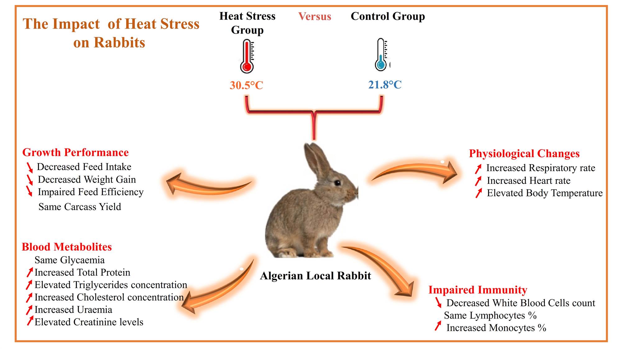
Effects of Heat Stress on Growth Performance, Carcass Traits, Physiological Components, and Biochemical Parameters in Local Algerian Growing Rabbits
Dahmani Y, Benali N, Saidj D, Chirane M, Ainbaziz H, and Temim S.
World Vet. J. 12(4): 405-417, 2022; pii:S232245682200051-12
DOI: https://dx.doi.org/10.54203/scil.2022.wvj51
ABSTRACT: Heat stress is a detrimental factor affecting the welfare of all livestock, especially rabbits, as they are sensitive to high temperatures. The current study investigated the effect of high ambient temperature on growth performance, slaughter traits, physiological indicators, and some hematological and biochemical parameters in Algerian local growing rabbits. A total of 48 local rabbits of both sexes (35 days old) were allotted into two groups (24 per group). The control group rabbits were exposed to an ambient temperature and humidity, averaging 21.8 ± 1.3°C and 51.7 ± 3.6%, respectively. Rabbits in the heat stress group were subjected to a warm ambient temperature and humidity of 30.5 ± 1.82°C and 65.5 ± 7.2%, respectively. The growth performance was measured and calculated from 35 to 91 days of age. Physiological indicators (rectal, skin, and ear temperatures, respiratory, and heart rates) were examined at 88 days of age. The carcass traits, blood metabolites, and hematological parameters of rabbits were measured and calculated at slaughter (92 days of age). The obtained results indicated a decrease in body weight, daily gain, and daily feed intake of rabbits in heat stress rabbits, compared to the control group. However, feed conversion ratio was significantly higher in the heat stress group, compared to the control. Heat stress group rabbits showed significantly higher blood metabolite levels, except the glycemia, which was similar in both groups. No significant effect of heat stress was found on the carcass yield, anterior, posterior, and intermediate parts of the carcass. However, the yield of the other components of the carcass (liver, kidney, peritoneal and inter-scapular fat) was significantly lower in the heat stress group. In the heat stress group, rectal, skin, and ear temperatures as well as heart and respiratory rates, were significantly higher than those of the control group. In the present experimental conditions, exposure of local rabbits to chronic heat stress could induce some changes to biological, physiological, and biochemical parameters leading to altered growth performance.
Keywords: Carcass yield, Growth performance, Heat stress, Local rabbit, Metabolic profile, Thermoregulation
[Full text-PDF] [Crossref Metadata] [Scopus] [Export from ePrint] [How to Cite]
 Research Paper
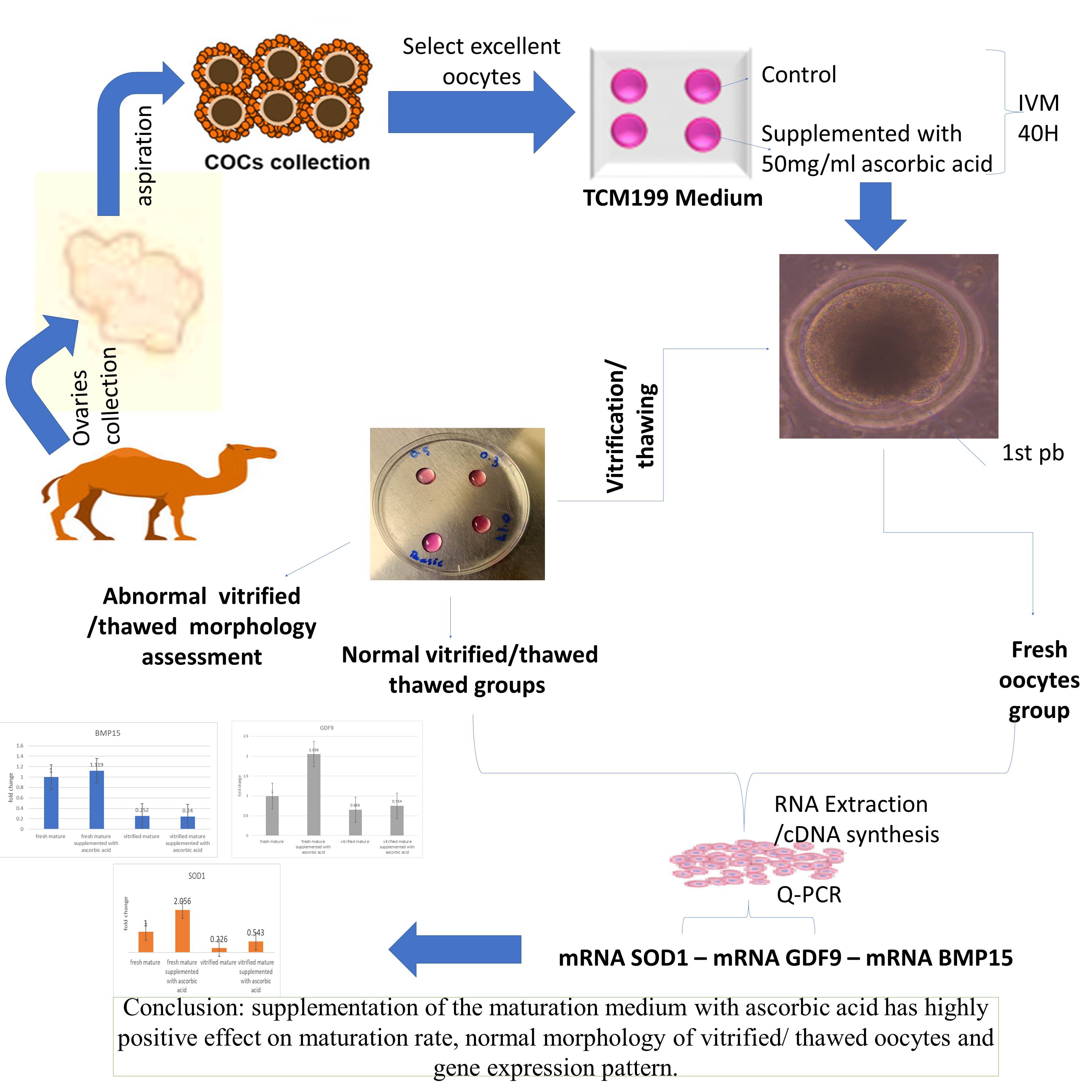
Effects of Ascorbic Acid on Maturation Rate, Morphology, and Gene Expression of Vitrified In Vitro Matured Dromedary Camel Oocytes
Kandil OM, Aboelwafa FB, Ismail EA, Kandeel SM, Ghanem N, and Gamal El-Din AE-K.
World Vet. J. 12(4): 418-429, 2022; pii:S232245682200052-12
DOI: https://dx.doi.org/10.54203/scil.2022.wvj52
ABSTRACT: In vitro embryo generation, cryopreservation, and embryo transfer are examples of assisted reproductive technologies that can be used to improve camel genetic performance and fertility. The aim of this study was to investigate the impact of ascorbic acid supplementation to in vitro maturation media on the maturation rate, morphology, and gene expression of fresh and vitrified in vitro matured dromedary camel oocytes. In the current study, 810 oocytes of excellent and good quality were in vitro matured in maturation medium (TCM-199 + 10 ug/ml follicle stimulated hormone + 10% fetal calf serum + 100 IU/ml Pregnant mare serum + 50 μg/ml gentamycin) without any additives to act as a control group (C) and with 50 μg/ml ascorbic acid group (AA) and incubation in a CO2 incubator (38.5 ̊C, 5% CO2, 20% O2 and 95% humidity) for 40 hours. In vitro matured dromedary camel oocytes with the first polar body (n = 210) in C group and AA group (n = 250) were placed in basic medium (BM) and then placed in vitrification solution1 (VS1) for one minute, followed by the transfer of oocytes to VS2 (double concentration of VS1, containing 20% Ethyl Glycol (EG) and +20% Dimethyl sulfoxide) for 30 seconds. Oocytes were then loaded into sterile 0.25 ml straws and stored in liquid nitrogen for 7-10 days. The normal fresh and vitrified /thawed in vitro matured dromedary camel oocytes were kept in RNA later at a -80°C freezer for gene expression analysis. The maturation rate of dromedary camel oocytes in the in vitro matured AA group was significantly higher than that of the C group. The percentage of normally recovered vitrified/thawed oocytes was higher in the in vitro matured with ascorbic acid (VAA) than in the control (VC) group. The expression pattern of the SOD1 gene and GDF9 gene was upregulated in fresh AA and VAA groups than in the fresh C and VC groups. The profile of the SOD1 gene was more abundant in the vitrified/thawed oocytes VAA group than in the VC group. All vitrified/thawed groups, whether control or ascorbic acid supplemented, had lower levels of SOD1, GDF9, and BMP15 expression, compared to the fresh groups. In conclusion, the supplementation of the maturation medium with ascorbic acid has an increased maturation rate, and normal morphology of vitrified/ thawed oocytes which was linked with upregulation of SOD1, GDF9 genes expression.
Keywords: Dromedary camel, Gene expression, In vitro maturation, Morphology, Vitrification
[Full text-PDF] [Crossref Metadata] [Scopus] [Export from ePrint] [How to Cite]
 Research Paper
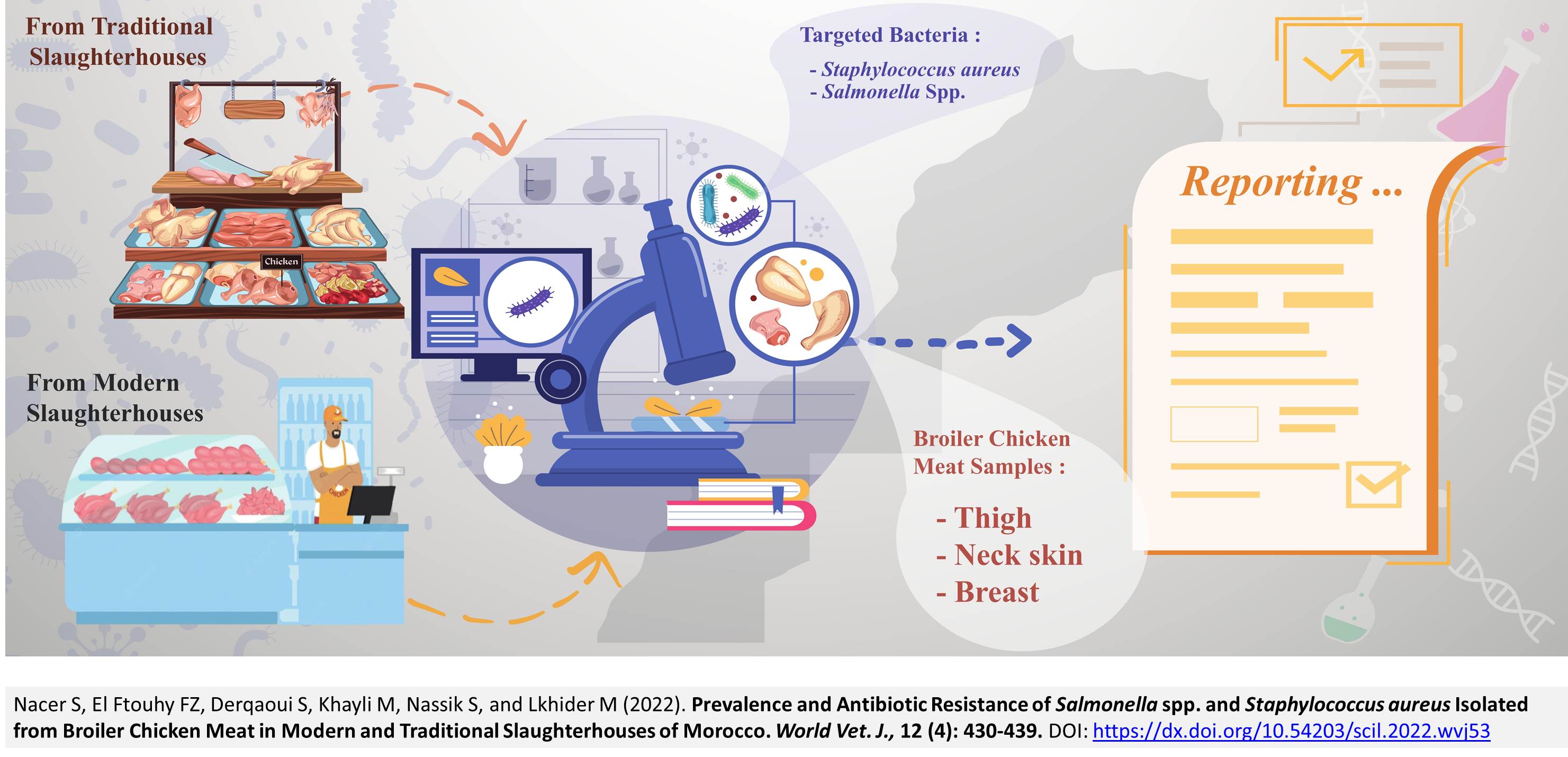
Prevalence and Antibiotic Resistance of Salmonella spp. and Staphylococcus aureus Isolated from Broiler Chicken Meat in Modern and Traditional Slaughterhouses of Morocco
Nacer S, El Ftouhy FZ, Derqaoui S, Khayli M, Nassik S, and Lkhider M.
World Vet. J. 12(4): 430-439, 2022; pii:S232245682200053-12
DOI: https://dx.doi.org/10.54203/scil.2022.wvj53
ABSTRACT: Handling and consuming contaminated meat can lead to food poisoning and the acquisition of antibiotic resistance genes. Staphylococcus aureus (S. aureus) and Salmonella spp. are the most isolated bacteria from broiler chicken meat, leading to serious foodborne diseases. The present study aimed to evaluate the presence and antibiogram profiles of Salmonella spp. and S. aureus strains in poultry meat purchased from modern and traditional poultry slaughterhouses in Morocco. Foodborne pathogens, such as Salmonella spp. and S. aureus, were isolated from poultry meat using standard methods and then confirmed by biochemical tests (coagulase, catalase, oxidase, motility and API 20E for further biochemical identification) and an immunological test (serotyping test). The antibiogram of the isolates was determined using the agar diffusion method and interpreted according to the criteria of performance standards for antimicrobial susceptibility testing of the Clinical and Laboratory Standards Institute, Wayne, Pennsylvania, USA. A total of 540 poultry meat samples were collected and treated (360 poultry meat samples from traditional slaughterhouses and 180 poultry meat samples from modern slaughterhouses), out of which 15.92% were S. aureus positive and 7.40% were Salmonella spp. positive. In traditional poultry slaughterhouses, the prevalence rates of Salmonella spp. and S. aureus were 11.11% and 20.55%, respectively. In contrast, Salmonella spp. was not detected in poultry samples of modern poultry slaughterhouses, and the prevalence of S. aureus was 6.66%. All S. aureus and 97% of Salmonella spp. isolates were found resistant to at least one antibiotic, while 86% of S. aureus and 30% of Salmonella spp. showed resistance to more than three antibiotics. The obtained results of the present study confirmed that broiler chicken meat purchased from traditional poultry slaughterhouses was mainly contaminated by Salmonella spp. and S. aureus, indicating a major public health risk in Morocco. Therefore, considerable efforts should be made to apply appropriate hygiene practices.
Keywords: Antimicrobial resistance, Broiler chicken meat, Modern slaughterhouses, Salmonella, Prevalence, Staphylococcus aureus, Traditional slaughterhouses
[Full text-PDF] [Crossref Metadata] [Scopus] [Export from ePrint] [How to Cite]
 Research Paper
Effects of Amphora Algae on Productive Performance and Immune Response of Broiler Chickens
El-Kaiaty AM, Elsherif HMR and Abdelaziz YM.
World Vet. J. 12(4): 440-448, 2022; pii:S232245682200054-12
DOI: https://dx.doi.org/10.54203/scil.2022.wvj54
ABSTRACT: Microalgae, especially Amphora coffeaeformis (A. coffeaeformis), are introduced to poultry diets, mainly as a rich source of polyunsaturated fatty acids (PUFAs), α-linolenic acid, eicosapentaenoic (EPA (and docosahexaenoic (DHA). This study aimed to investigate the effect of dietary supplementation of A. coffeaeformis on broiler chickens’ productive performance, physiological status, and immune response. A total of 180 (Ross 508) broiler chickens aged one day were wing banded and randomly divided into three treatments and a control group according to the form of A. coffeaeformis, with 45 chickens each. Each treatment had three replicates (15 chickens for each replicate). Chickens from the three treatments were fed a diet supplemented with A. coffeaeformis algae at levels of 0.15, 0.45, and 0.75% of the diet from the first week to the fifth weeks of age. The obtained results indicated a significant difference in live body weight (LBW), body weight gain (BWG), and growth rate (GR) at the different experimental periods due to the effects of A. coffeaeformis treatments compared to the control group. Chickens fed basal diet and diet with A. coffeaeformis at levels of 0.45%, and 0.75% significantly increased LBW, BWG, and GR% at all intervals (1-3), (3-5), and (1-5) weeks of age compared to A. coffeaeformis algae at levels of 0.15%. Chickens fed a diet supplemented with A. coffeaeformis 0.45% and AC 0.75% recorded higher plasma total protein insignificantly, albumin significantly, at five weeks of age compared to the other A. coffeaeformis treatments and control group. Moreover, the lower levels of plasma triglycerides, total cholesterol, LDL, and significantly higher levels of plasma HDL were found at a basal diet supplemented by A. coffeaeformis 0.15% and the control group. Also, AC 0.15% and A. coffeaeformis 0.45% recorded insignificantly lower plasma levels of Glutathione and Superproxedase (58.55 and 71.43 mg/l, respectively) when compared with other A. coffeaeformis treatments and control group. Dietary supplementation of chickens’ feed with A. coffeaeformis microalgae can promote the proliferation of beneficial bacteria (microbiota).
Keywords: Amphora coffeaeformis, Antioxidative status, Broiler chickens, Blood parameter, Immune response, Microalgae
[Full text-PDF] [Crossref Metadata] [Scopus] [Export from ePrint] [How to Cite]
 Research Paper
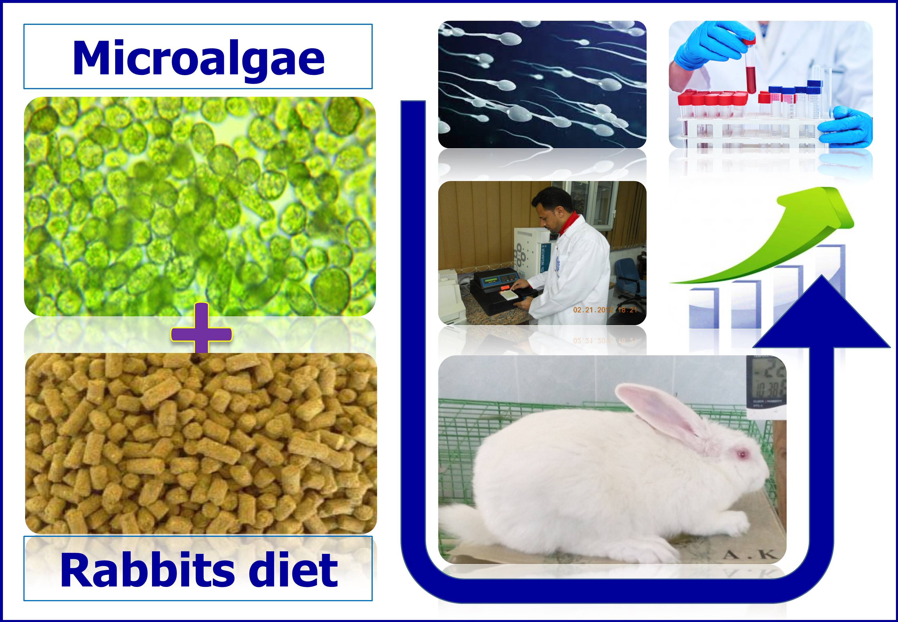
Semen Characteristics and Blood Metabolites of Hi-Plus Buck Rabbits Fed on Microalgae Nannochloropsis oculata Meal during the Summer Season
El-Hawy AS, El-Bassiony MF, Abd El-Hamid IS, Shedeed HA, Fouda WA, Ali SAM, Abd-Elazem RA, Morsy AS, and Emam KRS.
World Vet. J. 12(4): 449-458, 2022; pii:S232245682200055-12
DOI: https://dx.doi.org/10.54203/scil.2022.wvj55
ABSTRACT: Feeding tiny amounts of micro-algae meal to animals enhances animal physiology by improving immune response, disease resistance, and gut function, as well as enhancing anti-inflammatory and antibacterial protection, reproductive performance, feed conversion ratio, and weight gain. The purpose of this study was to identify the impact of dietary microalgae meal (Nannochloropsis oculata) on physical semen quality, serum biochemical parameters, and oxidative status of Hi-Plus buck rabbits for 12 weeks during the summer. A total of 45, Hi-Plus buck rabbits aged 20-24 weeks were divided into three equally comparable experimental groups. Bucks in the first, second, and third groups were daily supplemented in their diets with 0% (control), 0.50% (T1), and 1.0% (T2) microalgae meal, respectively. Semen and blood samples were collected to evaluate semen quality traits and some serum biochemical constituents, and oxidative status, as well as serum triiodothyronine (T3) and testosterone (Ts) hormones concentrations. The obtained data revealed that dietary supplementation of Nannochloropsis oculata meal significantly improved most physical semen characteristics, including ejaculate volume, progressive sperm motility, semen pH value, sperm cell concentration, total sperm output, live sperm, and semen quality factor. Blood serum glucose, total proteins, and their fractions increased significantly in T1 and T2, compared with the control group, while total serum cholesterol and hepatic enzymes concentrations recorded a significant decrease in bucks supplemented with T1 and T2, compared with the control group. The total antioxidant capacity of serum significantly increased in both two levels of microalgae, compared with the control group. Serum T3 concentration significantly increased in both levels of dietary microalgae compared with the control group. In conclusion, dietary supplementation with Nannochloropsis oculata meal (1.0%) was advised to improve semen quality, serum constituents, and antioxidative status without any adverse effects on the liver and kidney functions of rabbits.
Keywords: Bucks rabbits, Microalgae, Semen quality, Serum metabolite
[Full text-PDF] [Crossref Metadata] [Scopus] [Export from ePrint] [How to Cite]
 Protocol
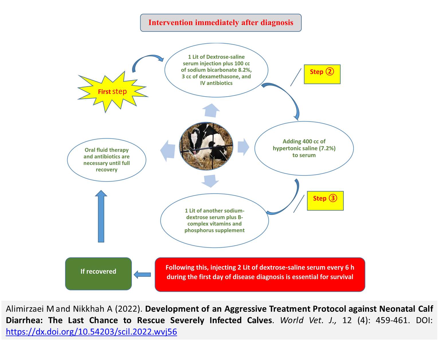
Development of an Aggressive Treatment Protocol against Neonatal Calf Diarrhea: The Last Chance to Rescue Severely Infected Calves
Alimirzaei M and Nikkhah A.
World Vet. J. 12(4): 459-461, 2022; pii:S232245682200056-12
DOI: https://dx.doi.org/10.54203/scil.2022.wvj56
ABSTRACT: Despite many efforts to control and treat neonatal calf diarrhea (NCD), it remains the primary cause of calf mortality in dairy herds worldwide. The objective of this article was to develop and discuss an empirical therapeutic protocol to save newborn calves with severe diarrhea. The pathophysiology of diarrhea has been well described previously. However, there is a significant gap between scientific findings and practical implementations. Reducing the number of calves with failure of passive transfer, regular sanitation of the calf environment, and optimal dry cow nutrition and management are fundamental measures in controlling diarrhea in commercial settings. As such, optimizing colostrum feeding management and improving ambiance hygiene are among the most important management practices to prevent calf diarrhea. Nonetheless, the occurrence of NCD would be unavoidable due to its multifactorial nature and pathophysiology. According to the degree of dehydration and general appearance of ill calves (e.g., degree of sunken eye and loss of suck reflex), NCD can be classified into mild to severe cases. Early diagnosis and treatment of both mild and severe cases could reduce pathogens shedding into the calf environment. Notably, diarrhea treatment needs profound scientific farm education and mentoring regarding the physiology of NCD. Since a variety of organisms, such as bacteria, viruses, and protozoa, may be responsible for NCD, it is evident that reliable diagnosis requires optimal sampling and laboratory analysis. However, waiting for laboratory results may waste the golden time of treatment. Therefore, rapid and decisive treatment would be mandatory, especially in severely infected calves or sepsis cases. Accordingly, an effective aggressive treatment protocol was developed and discussed in this article as the last chance to keep diarrheic calves alive.
Keywords: Aggressive treatment, Calf diarrhea, Dairy calf, Farm Management, Prevention
[Full text-PDF] [Crossref Metadata] [Scopus] [Export from ePrint] [How to Cite]
 Research Paper
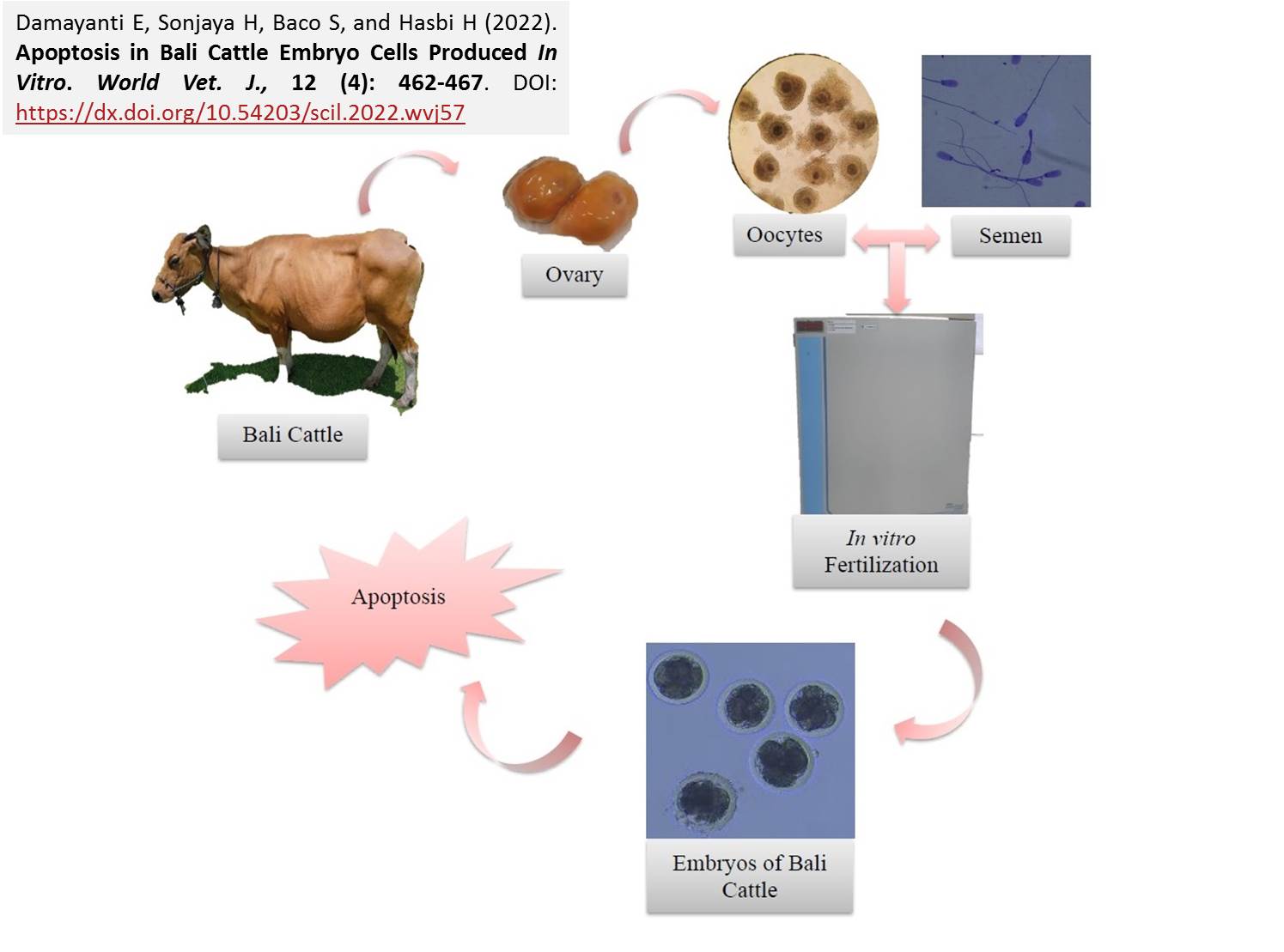
Apoptosis in Bali Cattle Embryo Cells Produced In Vitro
Damayanti E, Sonjaya H, Baco S, and Hasbi H.
World Vet. J. 12(4): 462-467, 2022; pii:S232245682200057-12
DOI: https://dx.doi.org/10.54203/scil.2022.wvj57
ABSTRACT: In vitro production of Bali cattle embryos still needs in-depth investigations to produce embryos suitable for transfer. The current study aimed to examine the level of cell apoptosis in Bali cattle embryos produced in vitro and at three stage of oocyte maturation, fertilization, and embryo culture. A total of 107 pairs of ovaries derived from slaughterhouses of Indonesia were collected. The used oocytes were grades A and B (Grade A had compact cumulus oocyte complex (COC) cells surrounded by five or more layers of cumulus cells, and grade B had a non-compact COC and a dark cytoplasm with complements from the complete radiata corona but surrounded by no more than five layers of cumulus cells). Fertilization of oocytes was done using the semen of a Bali bull. Bali cattle semen was frozen in straw semen for 5 minutes at 1500 rpm twice, then the supernatant and spermatozoa were separated and equilibrated for 30 minutes. Fertilization lasted for 5-6 hours in the incubator. Then, oocyte culture was carried out using CR1aa media and evaluated at 48 hours post-insemination (hpi). The result of the current study showed that the development of Bali cattle embryos produced in vitro after 48 hours of culture included 2 cells (31.91%), 4 cells (32.97%), 8 cells (24.46%), and 16 cells (10.63%). The percentage of embryos containing at least one nucleus exhibiting Terminal dUTP nick-end labeling (TUNEL) characteristics of apoptosis entailed 28.33% (2 cells), 41.93% (4 cells), 43.48% (8 cells), and 50% (16 cells). The division ability of embryos aged 48 hpi consisted of 2, 4, 8, and 16 cells. In conclusion, apoptosis in Bali cattle began to be detected in the two-cells stage. The sooner a cell undergoes apoptosis, the lower the level of the cell’s ability to develop further.
Keywords: Apoptosis, Bali cattle, Embryo, In vitro, cell cleavage
[Full text-PDF] [Crossref Metadata] [Scopus] [Export from ePrint] [How to Cite]
Previous issue | Next issue | Archive
![]() This work is licensed under a Creative Commons Attribution 4.0 International License (CC BY 4.0).
This work is licensed under a Creative Commons Attribution 4.0 International License (CC BY 4.0).


