
Previous issue | Next issue | Archive
![]() Volume 16 (2); June 2026
Volume 16 (2); June 2026
 Research Paper
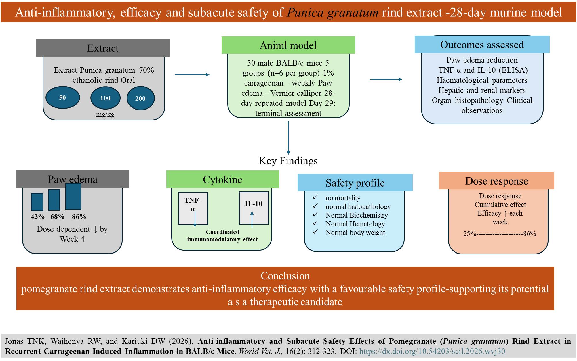
Anti-inflammatory and Subacute Safety Effects of Pomegranate (Punica granatum) Rind Extract in Recurrent Carrageenan-Induced Inflammation in BALB/c Mice
|
|
Jonas TNK, Waihenya RW, and Kariuki DW.
World Vet. J. 16(2): 312-323, 2026; pii:S232245682600030-16
DOI: https://dx.doi.org/10.54203/scil.2026.wvj30
ABSTRACT: Pomegranate (Punica granatum) contains a high level of polyphenols and has anti-inflammatory properties. However, there is limited available data on the safety and effectiveness of pomegranate use during prolonged inflammatory conditions. The present study aimed to evaluate the anti-inflammatory properties and subacute toxicity profile of a 70% ethanolic extract of Punica granatum (P. granatum) rind in carrageenan-induced mice. A total of 30 male mice, aged five weeks and weighing 22-23 grams, were randomly assigned to five groups of six mice each. The study included a negative control group that received normal saline, a positive control group that received meloxicam at 2 mg/kg, and three treatment groups that received P. granatum rind extract at doses of 50, 100, and 200 mg/kg orally over 28 days. Inflammation was induced once weekly in all experimental groups by 1% carrageenan injection into the right hind paw, after which the edema was measured with a digital vernier caliper. The toxicity of the pomegranate extract was assessed using hematological and biochemical profiles of hepatic and renal parameters. Tissue samples from the heart, spleen, kidney, lungs, and liver were collected on day 29. To assess the inflammation status, concentrations of tumor necrosis factor-alpha (TNF-α) and IL-10 were quantified using enzyme-linked immunosorbent assay. The P. granatum rind extract significantly reduced paw edema without mortality or observable toxicity, compared to the negative control group. Mice maintained their normal feeding habits, and body weight increased normally across all groups. Hematological parameters, hepatic, and renal markers were within the normal physiological range across all groups. Histopathological analysis revealed no abnormalities in extract-treated groups. Extract-treated groups demonstrated a dose-dependent elevation of IL-10, peaking at 200 mg/kg, and a reduction in TNF-α level across all doses, with the highest suppression observed at 100 mg/kg compared to the negative control. Daily oral administration of P. granatum rind extract for 28 days exhibited dose-dependent anti-inflammatory effects, as evidenced by reduced paw edema and modulation of cytokines. The absence of systemic toxicity across all tested doses suggested that P. granatum rind extract can be a safe therapeutic agent for treating inflammatory conditions.
Keywords: Anti-inflammatory, Biochemical marker, Carrageenan, Cytokine, Hematological parameter, Inflammation, Punica granatum
[Full text-PDF] [Crossref Metadata] [Scopus] [Export from ePrint]
 Research Paper
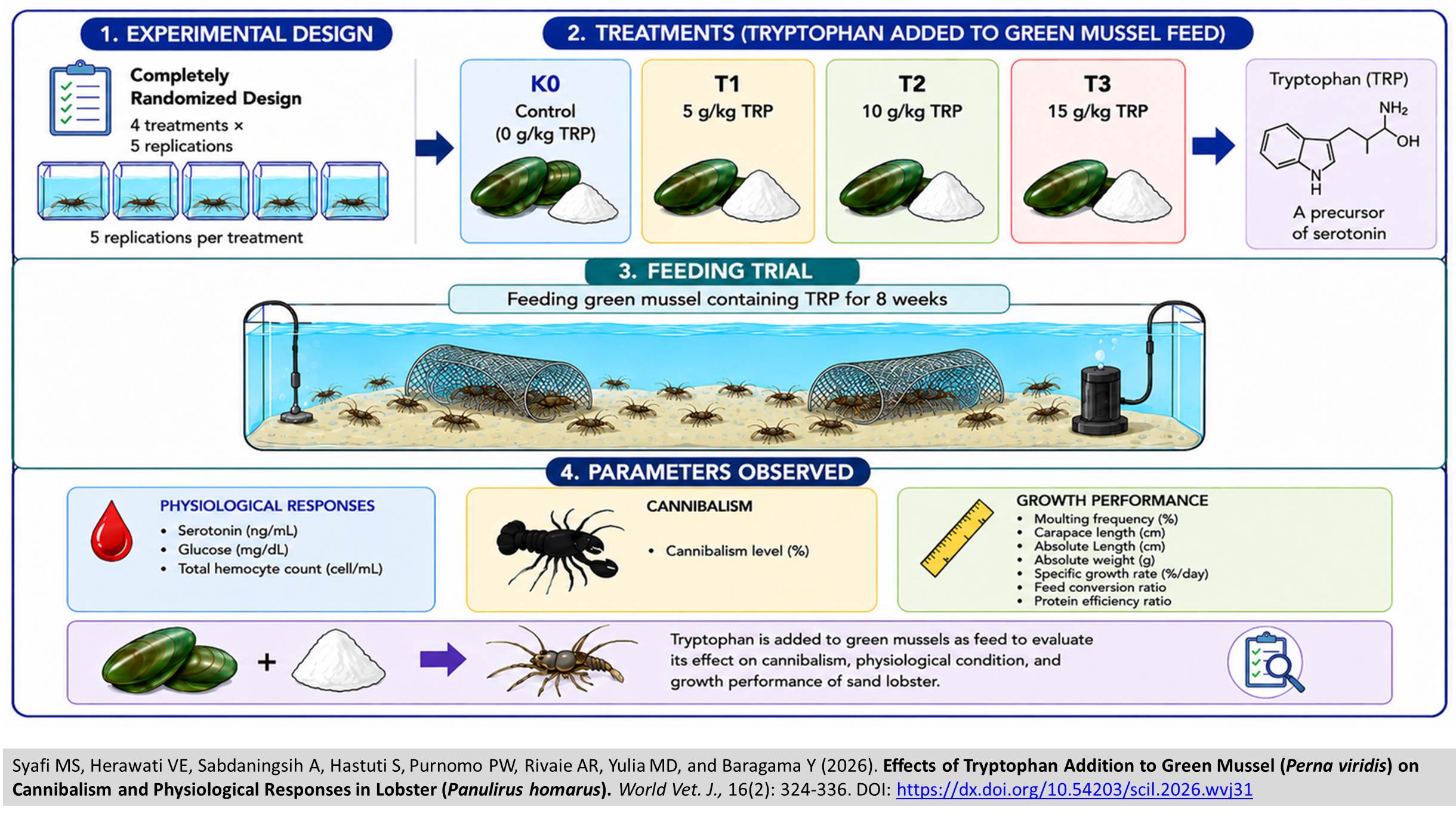
Effects of Tryptophan Addition to Green Mussel (Perna viridis) on Cannibalism and Physiological Responses in Lobster (Panulirus homarus)
|
|
Syafi MS, Herawati VE, Sabdaningsih A, Hastuti S, Purnomo PW, Rivaie AR, Yulia MD, and Baragama Y.
World Vet. J. 16(2): 324-336, 2026; pii:S232245682600031-16
DOI: https://dx.doi.org/10.54203/scil.2026.wvj31
ABSTRACT: The increasing demand for sand lobsters requires higher aquaculture productivity. However, sand lobster production is still constrained by high mortality due to cannibalism, a behavior in which individuals attack and consume conspecifics. One approach to suppress cannibalism is through supplementation of the amino acid tryptophan as a precursor to serotonin, which plays a role in regulating stress and aggressiveness. The present study aimed to determine the effectiveness and optimal formulation of tryptophan in green mussel feed on cannibalism, survival, and growth rate in sand lobsters. The study used a completely randomized design with four treatments and five replications. The treatments consisted of adding tryptophan to green mussels used as feed, including a control without tryptophan (K0), and supplementation at doses of 5 g/kg (T1), 10 g/kg (T2), and 15 g/kg (T3). Observed parameters included physiological responses, cannibalism levels, and growth. The results revealed that T3 treatment was most effective in increasing serotonin levels (89.45 ± 3.069 ng/mL) and survival rate (55.29 ± 5.26 %), and reducing cannibalism (7.06 ± 2.63 %), which caused lower glucose (14.19 ± 3.206 mg/dL) and total hemocyte count (81.00 ± 4.243 cell/mL) levels. However, the highest dose (T3; 15 g/kg) was less effective in enhancing growth performance, indicating that this level of supplementation was not optimal compared to the lower doses. In contrast, T1 (5 g/kg) resulted in relatively higher growth performance compared to the other treatments, as indicated by moulting frequency (1.40 ± 0.23%), carapace length (1.25 ± 0.15 cm), length increase (1.70 ± 0.12 cm), absolute weight (1.14 ± 0.35 g), specific growth rate (3.13 ± 0.46 %/day), feed conversion ratio (5.04 ± 0.93), and protein efficiency ratio (1.42 ± 0.18). However, in T1 (5 g/kg), serotonin (70.66 ± 4.165 ng/mL), survival rate (42.35 ± 4.92%), cannibalism (38.82 ± 3.22%), glucose (16.26 ± 3.352 mg/dL), and total hemocyte count (110.50 ± 9.192 cell/mL) were not as optimal as those observed in T3. Tryptophan supplementation revealed potential to enhance physiological condition, survival, and growth under the experimental conditions.
Keywords: Cannibalism, Growth, Lobster, Physiological, Tryptophan
[Full text-PDF] [Crossref Metadata] [Scopus] [Export from ePrint]
 Research Paper
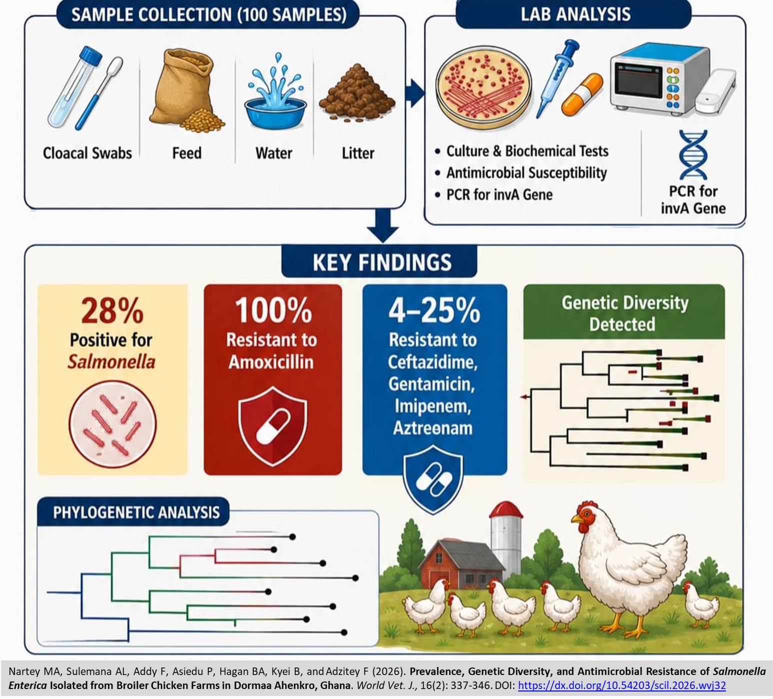
Prevalence, Genetic Diversity, and Antimicrobial Resistance of Salmonella Enterica Isolated from Broiler Chicken Farms in Dormaa Ahenkro, Ghana
|
|
Nartey MA, Sulemana AL, Addy F, Asiedu P, Hagan BA, Kyei B, and Adzitey F.
World Vet. J. 16(2): 337-346, 2026; pii:S232245682600032-16
DOI: https://dx.doi.org/10.54203/scil.2026.wvj32
ABSTRACT: Salmonella enterica (S. enterica) remains one of the most important bacterial agents linked to foodborne illness, particularly in regions with expanding poultry production. The present study aimed to investigate the occurrence, antimicrobial resistance, and genetic characteristics of S. enterica isolated from broiler chicken farms in Dormaa Ahenkro, Ghana. One hundred samples (cloacal swabs, feed, water, and litter) were collected from five farms and examined through culture, biochemical tests, antimicrobial susceptibility assays, and molecular confirmation of the invasion gene A (invA). Twenty-eight isolates (28%) were confirmed as Salmonella, all of which exhibited resistance to amoxicillin. Resistance to the antibiotics Ceftazidime, Gentamicin, Imipenem, and Aztreonam tested ranged from 4-25%. Nineteen isolates amplified the invA gene, and sixteen out of 19 submitted samples were successfully sequenced and confirmed as S. enterica with 93.85-99.80% identity. Phylogenetic and haplotype analyses demonstrated substantial genetic diversity, suggesting localized evolutionary divergence. The present findings highlight the presence of multidrug-resistant Salmonella within broiler production systems, emphasizing the need for improved biosecurity and responsible antimicrobial use to safeguard public health. Broiler chicken farms serve as an essential food source in many Ghanaian communities, yet they can harbour bacteria that pose risks to consumers. It was observed that Salmonella contamination in birds and their surroundings in Dormaa Ahenkro. Notably, Salmonella strains seen in these studies were also resistant to commonly used antibiotics, underscoring the need for stronger hygiene practices, prudent antibiotic use, and consistent monitoring to limit the spread of drug-resistant strains from farms to the wider population. Improving control measures will enhance food safety and support the sustainability of poultry production in Ghana.
Keywords: Antimicrobial resistance, Broiler chicken, Molecular characterization, Salmonella, One Health
[Full text-PDF] [Crossref Metadata] [Scopus] [Export from ePrint]
 Research Paper
Histopathological Insights into Purkinje Cell Responses of Maternal and Fetal Cerebellum in Mice: Electromagnetic Wave Exposure and Protective Role of Cactus (Opuntia spp.)
|
|
Armalina D, Susilaningsih N, Sutanto H, and Sunarno.
World Vet. J. 16(2): 347-359, 2026; pii:S232245682600033-16
DOI: https://dx.doi.org/10.54203/scil.2026.wvj33
ABSTRACT: The rapid expansion of wireless technologies has significantly increased human exposure to electromagnetic fields (EMFs), raising concerns about potential effects on reproductive and developmental health. Opuntia cochenillifera (O. cochenillifera), a cactus rich in mucilage and mineral oxides, has potential as a bio-based material for reducing the intensity of electromagnetic waves (EMWs) through dielectric and magnetic interactions. The present study aimed to evaluate the effects of EMW exposure on Purkinje cell morphology in maternal and fetal cerebellum and to assess the protective potential of distinct O. cochenillifera formulations. A total of 42 pregnant BALB/c mice were randomly divided into six groups, including healthy mice as the positive control group, EMW-exposed mice as the negative control group, and four treatment groups. The treatment groups were subjected to EMW exposure alongside the administration of either fresh cactus, dried cactus gel, or powdered cactus (3 g each). The EMW exposure was administered at a specific absorption rate of 1.74 W/kg throughout gestation. Cerebellar tissues were collected on day 20 for histological analysis and characterized by X-ray fluorescence and Fourier-transform infrared spectroscopy. Histological findings demonstrated that EMW exposure disrupted Purkinje cell alignment and decreased Purkinje cell counts in the negative control group. In contrast, all treatment groups demonstrated preserved cellular structure and morphology comparable to that of the healthy mice. Quantitative analysis confirmed significantly higher cell counts in treated groups than in the negative control group. The current findings indicated that EMW exposure adversely affected cerebellar development, while O. cochenillifera exhibited protective effects, supporting its potential as a natural EMF-attenuating material.
Keywords: Cactus, Cerebellum, Electromagnetic wave, Histopathology, Purkinje cell
[Full text-PDF] [Crossref Metadata] [Scopus] [Export from ePrint]
 Research Paper
Antifungal Activity of Crude Solanum incanum Fruit Extracts Against Trichophyton mentagrophytes and Exploratory Analysis of Virulence-Associated Gene Expression
|
|
Deta P, Ogoti P, Kiboi D, and Bii C.
World Vet. J. 16(2): 360-372, 2026; pii:S232245682600034-16
DOI: https://dx.doi.org/10.54203/scil.2026.wvj34
ABSTRACT: Dermatophyte infections in humans and animals are a significant public health concern, and increasing resistance to antifungal medicines highlights the need to explore alternative therapeutic agents. The present study aimed to preliminarily assess the in vitro antifungal activity of crude extracts from Solanum incanum (S. incanum) fruit against Trichophyton mentagrophytes (T. mentagrophytes). Solanum incanum fruits were extracted with hexane, ethyl acetate, methanol, and water. The levels of phenolic, flavonoid, and alkaloid compounds were measured, and antifungal activity was assessed using disc diffusion, minimum inhibitory concentration (MIC), and minimum fungicidal concentration (MFC) assays. Methanol and aqueous extracts exhibited the highest phenolic contents (~19 mg Gallic Acid Equivalents/g extract) and demonstrated significant antifungal activity, producing inhibition zones of 14.46 mm and 8.57 mm, respectively, at a concentration of 5 mg/mL. However, terbinafine demonstrated marginally greater inhibitory activity (15.02 mm) at a lower concentration of 0.1 mg/mL. The present findings indicated that using methanol and aqueous extracts of S. incanum fruit was associated with lower transcript levels of the targeted virulence-related genes (SUB3 and StuA). The current findings provided preliminary evidence regarding the antifungal activity of S. incanum fruit extract against T. mentagrophytes at a concentration of 5 mg/mL, indicating its potential as an antifungal agent.
Keywords: Antifungal activity, Bioactive compound, Dermatophyte, Gene expression, Medicinal plant
[Full text-PDF] [Crossref Metadata] [Scopus] [Export from ePrint]
 Research Paper
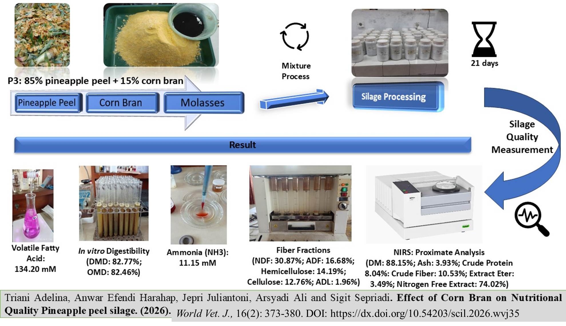
Effects of Corn Bran on the Nutritional Quality of Pineapple Peel Silage
|
|
Adelina T, Harahap AE, Juliantoni J, Ali A, and Sepriadi S.
World Vet. J. 16(2): 373-380, 2026; pii:S232245682600035-16
DOI: https://dx.doi.org/10.54203/scil.2026.wvj35
ABSTRACT: Combining pineapple peel waste with a specific level of corn bran produces high-quality silage that meets the nutritional needs of ruminants. The present study aimed to evaluate the effectiveness of incorporating corn bran in improving the physical properties and nutritional quality of pineapple peel silage for ruminant feeding in tropical regions. The silage treatment was conducted using a completely randomized design with four treatment groups and five replications. The experimental diets consisted of four levels of pineapple peel and corn bran, including 100% of pineapple peel with no bran (P0), 95% of pineapple peel with 5% of bran (P1), 90% of pineapple peel with 10% of bran (P2), and 85% of pineapple peel with 15% of bran (P3). Each treatment group received 5% of molasses as an additive. Silage quality was analyzed using near-infrared spectroscopy. To evaluate rumen fermentation and digestibility, the in vitro Conway method was utilized, employing rumen fluid obtained from fistulated Holstein-Friesian bulls. Pineapple peel silage containing different corn starches exhibited a higher pH than the control group. Group P3 significantly produced higher crude protein (CP) and nitrogen-free extract (NFE) levels than the other treatment groups. Increasing the amount of corn bran in group P3 increased ammonia (NH3) production by 11.15 mM and total volatile fatty acids (VFA) by 134.20 mM compared to the control group. Group P3 exhibited the highest digestibility coefficients, with dry matter and organic matter values reaching 82.77% and 82.46%, respectively. The feed mixture comprising 85% pineapple peel and 15% corn bran is recommended for ruminants in tropical regions due to its superior overall nutritional quality. This feed mixture elevated CP, NFE, NH3 production, total VFA, dry matter, and organic matter.
Keywords: Corn bran, Digestibility, Rumen, Tropical region
[Full text-PDF] [Crossref Metadata] [Scopus] [Export from ePrint]
 Research Paper
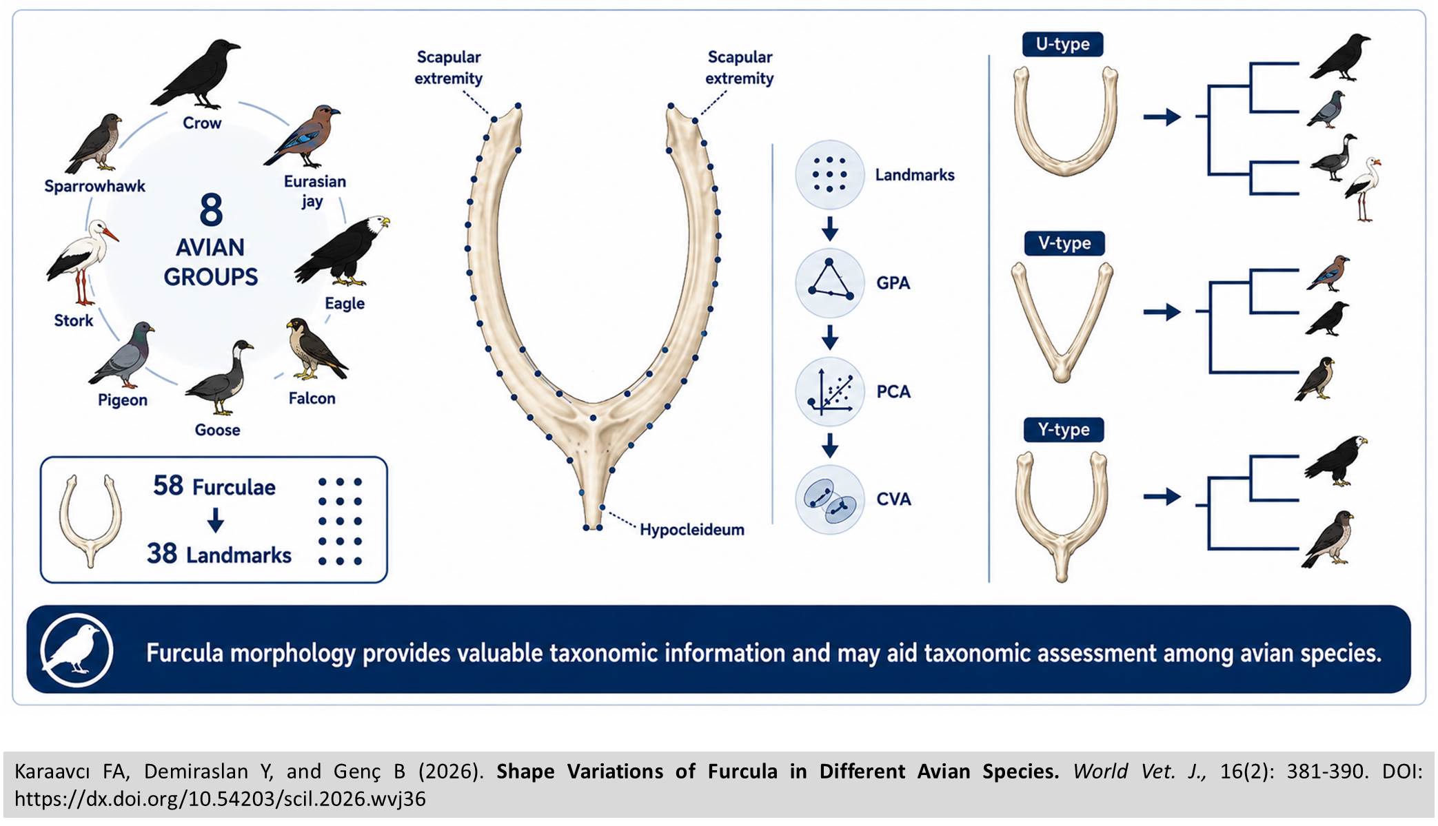
Shape Variations of Furcula in Different Avian Species
|
|
Karaavcı FA, Demiraslan Y, and Genç B.
World Vet. J. 16(2): 381-390, 2026; pii:S232245682600036-16
DOI: https://dx.doi.org/10.54203/scil.2026.wvj36
ABSTRACT: The furcula is a distinctive bone in avian species formed by the fusion of the left and right clavicles into a single structure. By acting as a spring during flight, the furcula adjusts the distance between the wing and the shoulder joint, contributing significantly to avian flight. The present study aimed to analyze furcular shapes across several bird species and investigate how these differences influence avian taxonomy. A total of 58 furculae collected from wild adult birds found dead or that died during treatment between 2023 and 2024 were examined. Samples consisted of seven crows, eight Eurasian jays, two eagles, one falcon, 19 pigeons, two storks, 10 sparrowhawks, and nine geese. Furculae were dissected and morphologically examined for overall shape, hypocleideum morphology, and the presence of pneumatic foramina. Morphological examination revealed that the furcula was V-shaped in the crow, Eurasian jay, goose, and pigeon, U-shaped in the eagle, falcon, and sparrowhawk, and Y-shaped in the stork. Geometric morphometric analysis was performed with 38 landmarks. Geometric morphometric analysis revealed that shape variation was primarily concentrated at the scapular ends of the furcula and secondarily in the hypocleideum region. Crow and Eurasian jay samples exhibited relatively similar shape patterns, whereas significant differences were detected among species groups in the canonical variate analysis. The current findings demonstrated species-specific variation in furcular morphology and provided valuable information for avian taxonomy and the functional interpretation of flight-related skeletal adaptations.
Keywords: Avian, Furcula, Geometric morphometry, Morphology, Shape
[Full text-PDF] [Crossref Metadata] [Scopus] [Export from ePrint]
 Research Paper
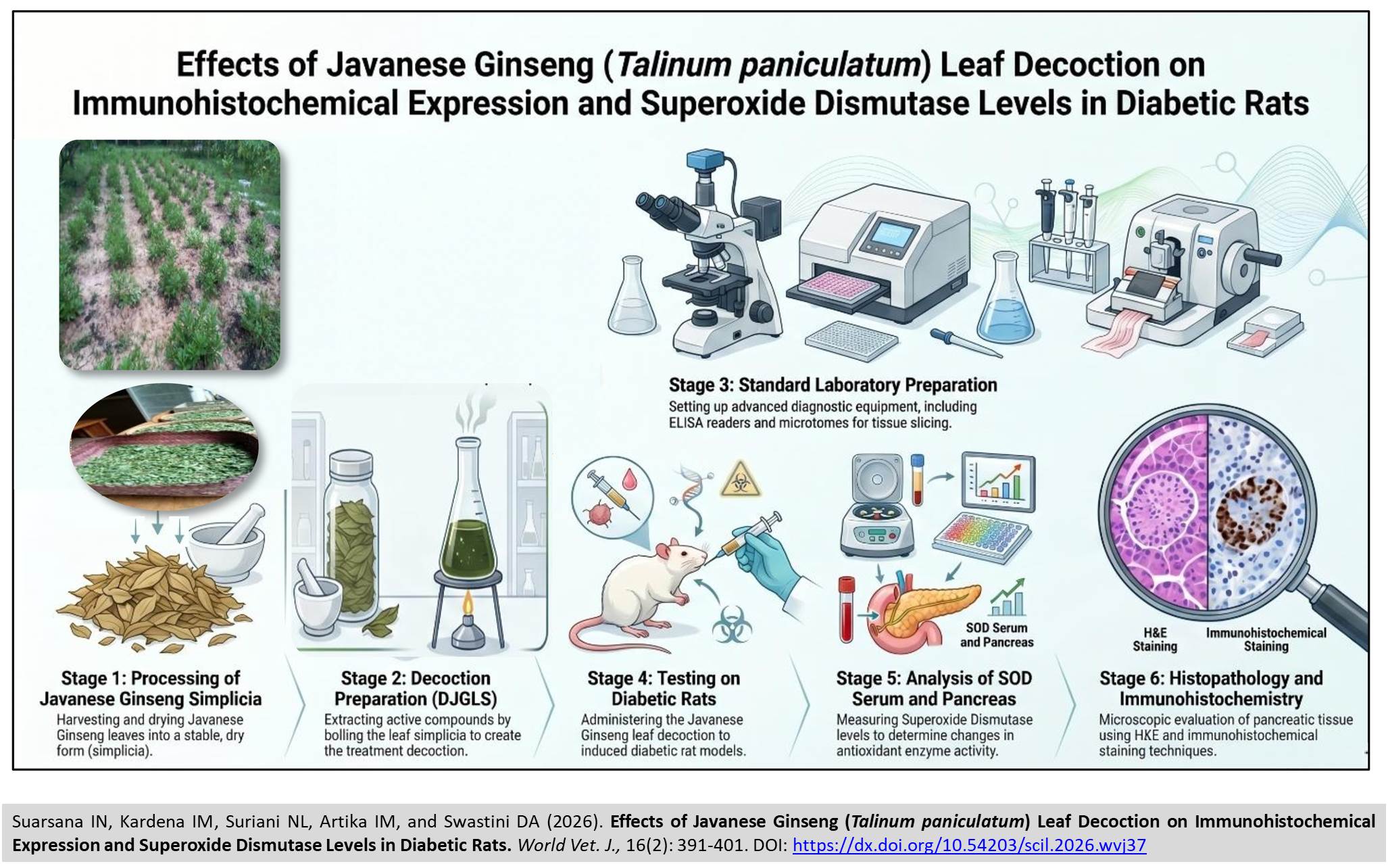
Effects of Javanese Ginseng (Talinum paniculatum) Leaf Decoction on Immunohistochemical Expression and Superoxide Dismutase Levels in Diabetic Rats
|
|
Suarsana IN, Kardena IM, Suriani NL, Artika IM, and Swastini DA.
World Vet. J. 16(2): 391-401, 2026; pii:S232245682600037-16
DOI: https://dx.doi.org/10.54203/scil.2026.wvj37
ABSTRACT: Java Ginseng has long been used by Indonesian communities as a medicinal plant and is traditionally consumed as an alternative therapy, even in managing Diabetes Mellitus (DM). The present study aimed to evaluate the effects of the decoction of Javanese ginseng leaf simplicia (DJGLS) on blood glucose levels, serum superoxide dismutase (SOD), the levels and expression intensity of SOD, and pancreatic histopathology in DM rats. Twenty adult male rats were randomly assigned to four groups. The normal control group (GJ-0, n = 5), the DM group (GJ-1, n = 5) received a placebo, and the DM groups (GJ-2, n = 5 and GJ-3, n = 5) were administered a DJGLS (100 g/100 mL, w/v powder simplicia), at volume of 0.5 mL and 1 mL/100 g body weight, respectively for 21 consecutive days. Blood glucose levels were measured using a glucometer. Serum and pancreatic SOD levels were determined using an ELISA kit. Pancreatic histopathology was evaluated using hematoxylin and eosin staining, while pancreatic SOD expression was assessed by immunohistochemical staining. In addition, phytochemical screening was performed on the Javanese ginseng leaf simplicia powder. The present study demonstrated that the phytochemical analysis of Java Ginseng leaf simplicia powder revealed the presence of alkaloids, flavonoids, saponins, tannins, and steroids. DJGLS treatment reduced blood glucose levels and serum SOD levels in diabetic rats, with a significant reduction observed in the GJ-3 group compared with the GJ-1. Administration of DJGLS in the GJ-2 and GJ-3 groups significantly increased pancreatic SOD levels compared with the GJ-1. The DJGLS treatment in the GJ-2 and GJ-3 groups demonstrated restorative effects, as evidenced by the recovery of the Islets of Langerhans area and increased intensity of pancreatic SOD expression compared with the GJ-1. In conclusion, DJGLS treatment was able to reduce blood glucose levels and serum SOD levels, although these values had not fully returned to normal conditions, and it increased pancreatic SOD levels. The DJGLS indicated potential for promoting recovery of the pancreatic Islets of Langerhans area.
Keywords: Decoction, Diabetes, Immunohistochemistry, Javanese ginseng, Superoxide dismutase
[Full text-PDF] [Crossref Metadata] [Scopus] [Export from ePrint]
 Research Paper
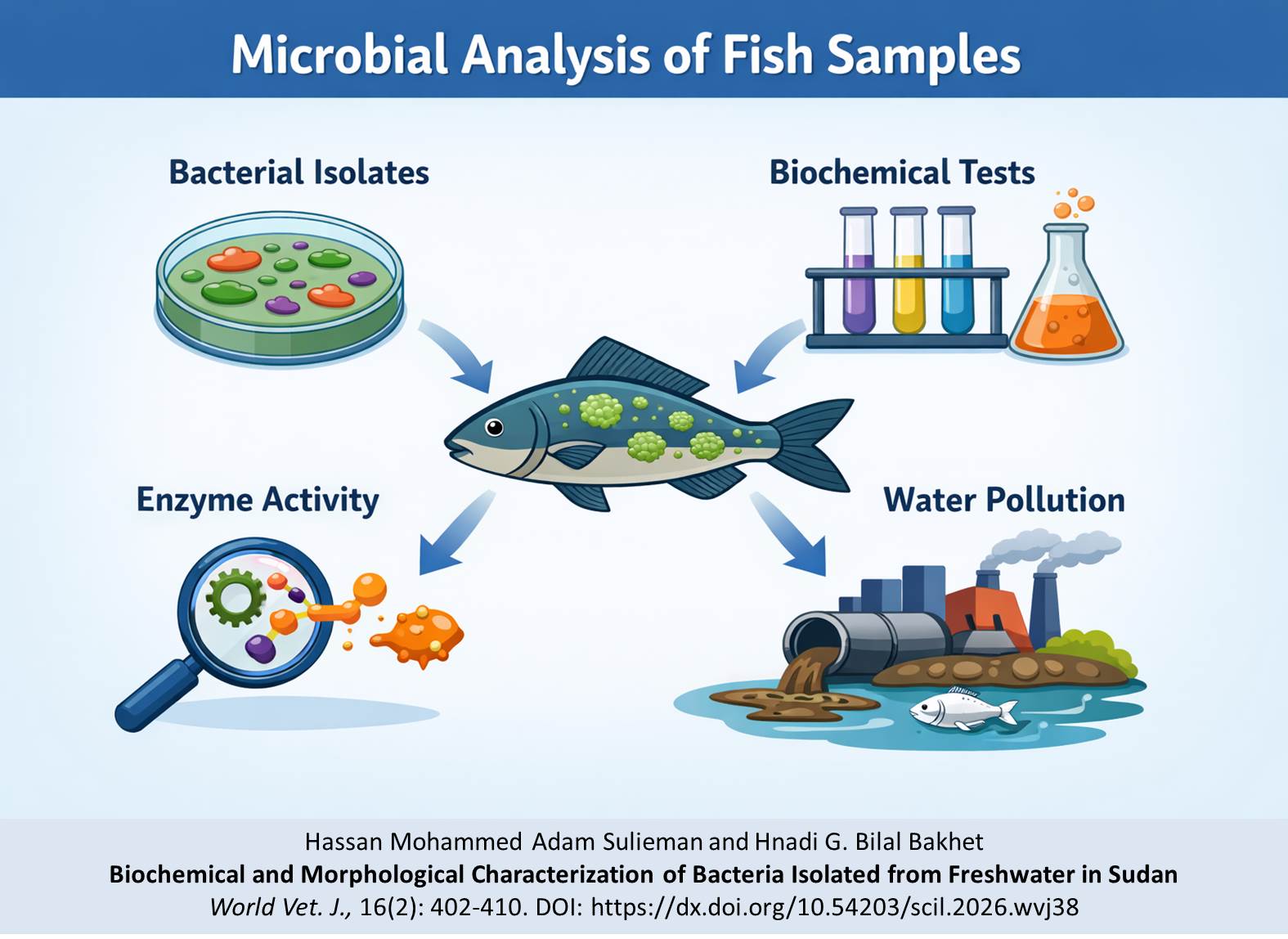
Biochemical and Morphological Characterization of Bacteria Isolated from Freshwater in Sudan
|
|
Sulieman HMA and Bakhet HGAB.
World Vet. J. 16(2): 402-410, 2026; pii:S232245682600038-16
DOI: https://dx.doi.org/10.54203/scil.2026.wvj38
ABSTRACT: The microbial quality of freshwater fish is a crucial indicator of both aquatic environmental health and food safety. The present study aimed to isolate and characterize bacteria from three common freshwater fish species, including Oreochromis niloticus, Clarias gariepinus, and Synodontis alberti, collected from three sites in Khartoum, Sudan. Six fish of each species were collected from each sampling location where the species were present. Total viable bacterial counts in fish gill and intestinal tissues obtained from Green Belt Sewage, Jebel Aulia fish landing, and Al Mourda fish market ranged from 1.2 × 10⁴ to 3.9 × 10⁵ CFU/g, with higher bacterial loads generally observed in intestinal tissues compared to gills. The highest bacterial load (3.9 × 10⁵ CFU/g) was recorded in the intestine of Clarias species collected from Green Belt Sewage. The current results demonstrated a predominance of Gram-negative, rod-shaped bacteria in gill and intestinal tissue samples across all sampling sites. All isolates were catalase-positive and capable of fermenting glucose, indicating facultative anaerobic metabolism. Oxidase activity of bacterial isolates differed by location; fish samples from Al Murda Fish Market had the highest number of oxidase-positive isolates, suggesting the potential presence of Aeromonas and Pseudomonas species. Urease activity was predominantly observed in isolates of fish samples collected from Al Murda and Jebel Aulia, suggesting a greater risk of fish spoilage or pathogenicity. Notably, isolates from the Green Belt Sewage fish samples comprised Gram-positive coccus, potentially identified as Staphylococcus spp., underscoring the likelihood of anthropogenic contamination. The present results indicated that fish obtained from all sampling locations generally exhibited similar microbial communities. However, the differences in enzyme activity across fish from all sites likely reflected variations in environmental factors, sanitation practices, and potential public health risks.
Keywords: Bacterial isolate, Biochemical characterization, Enzyme activity, Fish, Microbial count
[Full text-PDF] [Crossref Metadata] [Scopus] [Export from ePrint]
 Research Paper
Monitoring of Calcium and Phosphorus in Ruminant Feed by Smartphone-Assisted Colorimetry
|
|
Suhubdy S, Dilaga SH, Noersidiq A, and Ahmadi A.
World Vet. J. 16(2): 411-417, 2026; pii:S232245682600039-16
DOI: https://dx.doi.org/10.54203/scil.2026.wvj39
ABSTRACT: Routine laboratory analysis for ruminant feed minerals is often unavailable in a field setting; therefore, rapid mineral monitoring is necessary. The present study aimed to evaluate a smartphone-assisted colorimetry method for estimating calcium and phosphorus in forage extracts and compare its performance with UV-Visible spectrophotometry. Standard solutions ranging from 1 to 5 ppm were prepared for smartphone-assisted colorimetry and UV-Visible spectrophotometry, and reagent-treated extracts from four forage species, including Pennisetum purpureum cv. Mott, Pennisetum purpupoides, Leucaena leucocephala, and Sesbania grandiflora were measured by a Genesys 10S spectrophotometer and by smartphone imaging under controlled lighting. Linear regression was used to develop calibration models, and method agreement was assessed using Pearson's r and mean absolute percentage error. Both methods indicated strong linearity for calcium and phosphorus. For the smartphone method, the coefficient of determination ranged from 0.9448 to 0.9974, compared to 0.9514 to 0.9610 for the UV-Vis method, with absolute differences between the two methods remaining below 0.5%. Smartphone-assisted colorimetry and UV-Visible spectrophotometry produced closely comparable estimates for each tested forage sample for the tested forages. Smartphone-assisted colorimetry provided accurate estimates of calcium and phosphorus in forage extracts, comparable to those produced by spectrophotometry, and demonstrated potential as a low-cost tool for routine screening of ruminant feed minerals.
Keywords: Calcium, Colorimetry, Forage, Nutrition, Phosphorus, Ruminant feed, Smartphone
[Full text-PDF] [Crossref Metadata] [Scopus] [Export from ePrint]
Previous issue | Next issue | Archive
![]() This work is licensed under a Creative Commons Attribution 4.0 International License (CC BY 4.0).
This work is licensed under a Creative Commons Attribution 4.0 International License (CC BY 4.0).