Previous issue | Next issue | Archive
![]() Volume 15 (1); March, 2025 [Booklet] [EndNote XML for Agris]
Volume 15 (1); March, 2025 [Booklet] [EndNote XML for Agris]
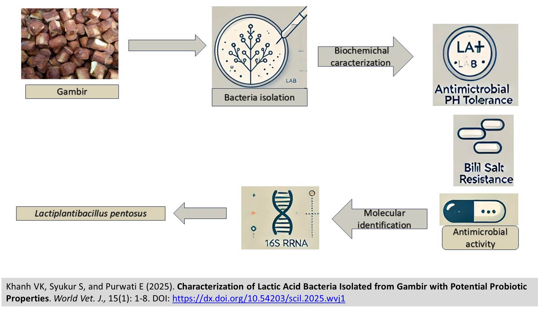
Characterization of Lactic Acid Bacteria Isolated from Gambir with Potential Probiotic Properties
Khanh VK, Syukur S, and Purwati E.
World Vet. J. 15(1): 1-8, 2025; pii:S232245682500001-15
DOI: https://dx.doi.org/10.54203/scil.2025.wvj1
ABSTRACT: Gambir is commonly used as a key ingredient in betel quid and is one of Indonesia's major agricultural commodities. West Sumatra is the primary production region, contributing approximately 80-90% of the country's total gambir production. This study aimed to investigate the potential of lactic acid bacteria (LAB) isolated from gambir, assessing their potential probiotic qualities using biochemical and molecular approaches. Biochemical characterization included catalase activity testing and fermentation pattern analysis, while molecular identification was carried out through the 16S rRNA gene sequence. The results revealed that the isolated LAB strains were Gram-positive, bacilli-shaped, catalase-negative, and exhibited hetero-fermentative behavior. Further biochemical analysis confirmed their ability to ferment a variety of sugars but not produce gas from glucose. Basic Local Alignment Search Tool analysis showed that the bacterial isolate from gambir, labeled with the sample code GM2, was closely related to Lactiplantibacillus pentosus, a species recognized for its probiotic potential. The isolates showed antimicrobial activity against common foodborne pathogens like Salmonella spp. and other pathogens, suggesting their potential use in food preservation. The present study also demonstrated the LAB isolates’ tolerance to low pH and bile salts, which are key attributes for probiotic candidates. Thus, the findings of the current study suggest that LAB from gambir possess promising characteristics for application in probiotic products and as biocontrol agents in food safety.
Keywords: 16s rRNA, Gambir, Lactic acid bacteria, Probiotic
[Full text-PDF] [Crossref Metadata] [Scopus] [Export from ePrint]
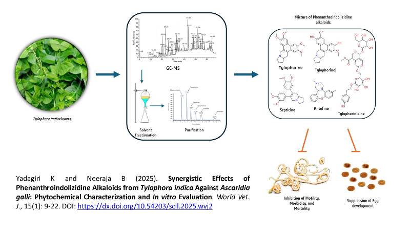
Synergistic Effects of Phenanthroindolizidine Alkaloids from Tylophora indica Against Ascaridia galli: Phytochemical Characterization and In vitro Evaluation
Yadagiri K and Neeraja B.
World Vet. J. 15(1): 9-22, 2025; pii:S232245682500002-15
DOI: https://dx.doi.org/10.54203/scil.2025.wvj2
ABSTRACT: The increasing resistance of helminths such as Ascaridia galli to conventional anthelmintics has necessitated the search for alternative treatments from natural sources. This study aimed to assess the anthelmintic properties of phenanthroindolizidine alkaloids derived from Tylophora indica leaves, a plant renowned for its medicinal value, including its traditional use in treating respiratory disorders, inflammation, and various infections. The leaves were subjected to sequential extraction with chloroform, ethanol, and water, which produced yields of 7.2%, 17.8%, and 11.2%, respectively. Phytochemical analysis revealed that the ethanol extract was rich in bioactive compounds, including significant amounts of alkaloids, flavonoids, phenols, and terpenoids. Quantitative analysis confirmed the ethanol extract's superiority, displaying the highest contents of phenolics (7.51 ± 0.62 mg/g), flavonoids (9.34 ± 1.63 mg/g), and alkaloids (17.65 ± 1.69 mg/g), underscoring its potential for various therapeutic applications. Further fractionation and High-Performance Liquid Chromatography (HPLC) purification isolated key phenanthroindolizidine alkaloids, including Tylophorinidine, Tylophorine, Septicine, Tylophorinol, and Antofine. Structural characterization via Nuclear Magnetic Resonance (NMR) and High-Resolution Mass Spectrometry (HRMS) validated these compounds. In vitro assays demonstrated significant dose-dependent anthelmintic activity against Ascaridia galli worms. Ethanol extracts exhibited the highest mortality rates, achieving 100% mortality within 24 hours at a concentration of 5 mg/mL. The mixture of all five alkaloids at 500 µg/mL showed a synergistic effect, leading to rapid and complete anthelmintic action. The egg embryonation assay further highlighted the efficacy of these alkaloids. The egg embryonation assay further demonstrated the potent efficacy of these alkaloids, with the mixture at 500 µg/mL inhibiting 92.67% of egg development, surpassing the positive control, i.e. piperazine citrate, which showed 87.25% inhibition. Among individual alkaloids, Tylophorinidine exhibited the highest inhibition of egg embryonation (80.76%), followed by Antofine (78.42%). These findings demonstrated the potent anthelmintic properties of phenanthroindolizidine alkaloids from Tylophora indica, particularly when used in combination (Tylophorinidine, Tylophorine, Septicine, Tylophorinol, and Antofine), compared to their individual effects. The study underscores the potential of these compounds as effective treatments for helminth infections and highlights the importance of further research to isolate specific mechanisms and optimize their therapeutic efficacy.
Keywords: Anthelmintic activity, Ascaridia galli, Phenanthroindolizidine alkaloids, Phytochemical analysis, Tylophora indica
[Full text-PDF] [Crossref Metadata] [Scopus] [Export from ePrint]
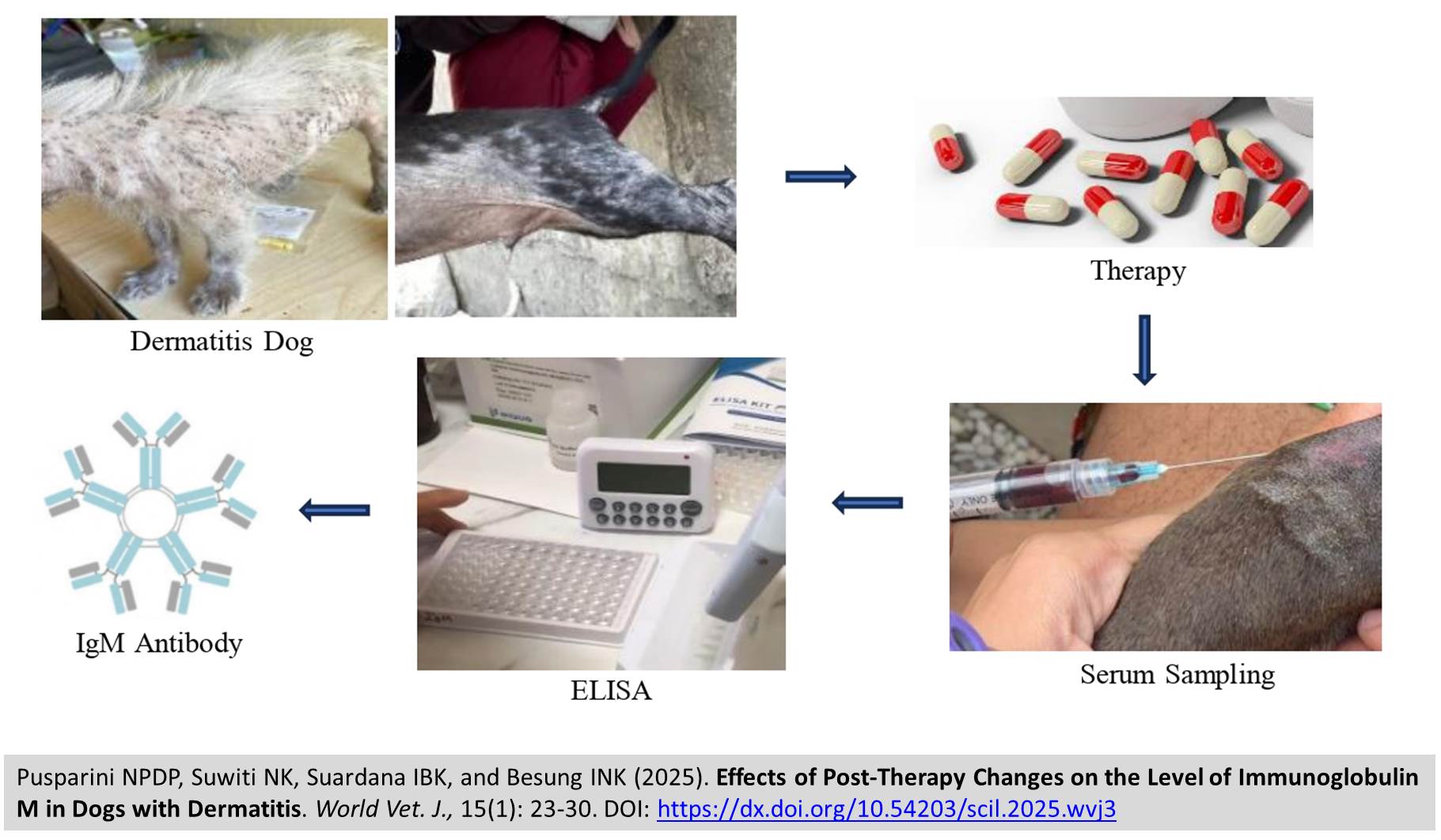
Effects of Post-Therapy Changes on the Level of Immunoglobulin M in Dogs with Dermatitis
Pusparini NPDP, Suwiti NK, Suardana IBK, and Besung INK.
World Vet. J. 15(1): 23-30, 2025; pii:S232245682500003-15
DOI: https://dx.doi.org/10.54203/scil.2025.wvj3
ABSTRACT: Dermatitis is an inflammation of the skin characterized by itching, hair loss, lesions, and redness. Various agents can cause dermatitis, including Sarcoptes scabiei, Demodex canis, and Microsporum canis. Animals experiencing dermatitis undergo internal changes in their bodies, particularly in the immune system. The presence of an infection is usually preceded by the appearance of Immunoglobulin M (IgM). This study aimed to determine the differences in IgM levels in dogs with dermatitis before therapy (pre-therapy) and after therapy (post-therapy), as well as the differences in IgM levels between dogs with mild and severe dermatitis. The study involved 40 local dogs, divided into two groups, including 20 dogs with mild dermatitis and 20 dogs with severe dermatitis. Serum sampling was conducted in two phases: the first phase was pre-therapy, and the second phase was 14 days after therapy (post-therapy). The therapy administered to dogs with mild dermatitis consisted of diphenhydramine HCl and ivermectin, while the therapy for dogs with severe dermatitis included diphenhydramine HCl, ivermectin, amoxicillin, and dexamethasone. Serum samples from the dogs were then tested using the Enzyme-Linked Immunosorbent Assay method. The results of the study revealed that serum IgM levels in dogs with mild and severe dermatitis did not show any significant difference. In dogs with mild dermatitis, serum IgM levels before therapy were not statistically different compared to those after therapy. However, in dogs with severe dermatitis, serum IgM levels before therapy were significantly higher compared to after therapy. The results of this study indicate that therapy can impact serum IgM levels in dogs with severe dermatitis, while it does not significantly affect these levels in cases of mild dermatitis.
Keywords: Dermatitis, Dog, Enzyme-linked immunosorbent assay, Immunoglobulin M, Ivermectin, Therapy
[Full text-PDF] [Crossref Metadata] [Scopus] [Export from ePrint]
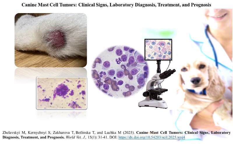
Canine Mast Cell Tumors: Clinical Signs, Laboratory Diagnosis, Treatment, and Prognosis
Zhelavskyi M, Kernychnyi S, Zakharova T, Betlinska T, and Luchkа M.
World Vet. J. 15(1): 31-41, 2025; pii:S232245682500004-15
DOI: https://dx.doi.org/10.54203/scil.2025.wvj4
ABSTRACT: Canine mast cell tumors, a tumor originating from mast cells involved in allergic reactions and inflammation, are among the most common skin tumors in dogs. The present study aimed to explore the clinical features, diagnostic approaches, and prognosis of canine mastocytomas through a case study. A 5-year-old male Akita, weighing 35.8 kg, was brought to the Doctor VET veterinary clinic in Kamianets-Podilskyi, Ukraine, for evaluation. Upon initial examination, the dog had a body temperature of 38.5°C, a heart rate of 74 beats per minute (bpm), and a respiratory rate of 28 breaths per minute, all of which were within normal physiological limits. The animal was alert and responsive and displayed no signs of systemic distress. A detailed physical examination revealed a tumor located 35.2 mm below the plantar surface of the tarsal joint (art. tarsi). The tumor was round, mobile, and surrounded by a thin fibrous capsule, with no signs of pain or discomfort during palpation. Cytological analysis showed a high-cellularity smear with numerous mast cells scattered throughout the field. These cells were round to oval in shape with abundant cytoplasm containing dense, basophilic to metachromatic granules. The hematological evaluation indicated a systemic inflammatory or immune response triggered by the tumor, as evidenced by neutrophilic leukocytosis (73.1%; 8.89×10⁹/L). Biochemical analysis revealed an elevated alkaline phosphatase activity level (4.45 μmol/L), suggesting systemic involvement. The tumor was surgically excised, ensuring complete removal with wide margins to minimize the risk of recurrence. Histological examination of the excised tissues confirmed a densely cellular neoplastic infiltrate composed predominantly of mast cells arranged in sheets and clusters. The mast cells displayed significant cellular and nuclear pleomorphism, characterized by moderate to marked anisocytosis and anisokaryosis. While no significant necrosis was observed, scattered apoptotic bodies were present, indicating ongoing cellular turnover. This case highlighted the critical importance of early diagnosis and comprehensive management of canine mastocytomas. Low-grade tumors often carry a favorable prognosis when treated promptly and appropriately. However, higher-grade or poorly differentiated tumors may require multimodal therapeutic approaches to achieve better outcomes.
Keywords: Canine, Diagnosis, Mast cell tumor, Mastocytoma, Skin tumor
[Full text-PDF] [Crossref Metadata] [Scopus] [Export from ePrint]
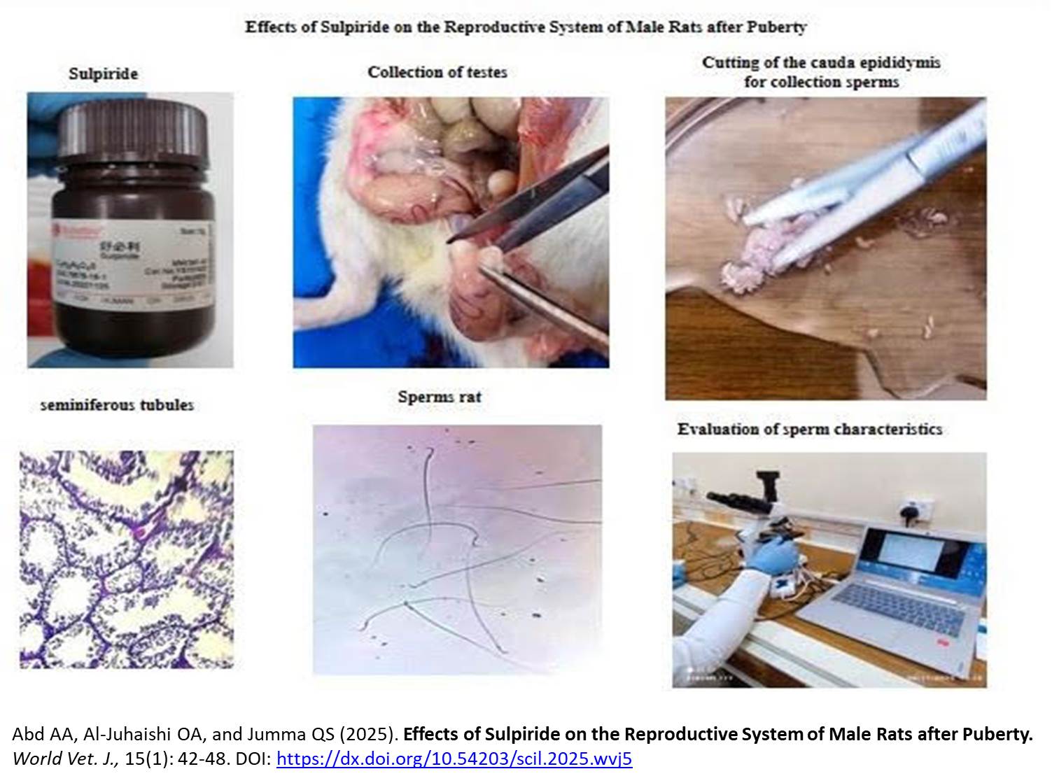
Effects of Sulpiride on the Reproductive System of Male Rats after Puberty
Abd AA, Al-Juhaishi OA, and Jumma QS.
World Vet. J. 15(1): 42-48, 2025; pii:S232245682500005-15
DOI: https://dx.doi.org/10.54203/scil.2025.wvj5
ABSTRACT: Sulpiride is an antipsychotic drug commonly used in humans to mitigate the effects of stress by selectively targeting central dopaminergic receptors. During male rat puberty, neurotransmitter systems, including the dopaminergic system, undergo significant development, playing a crucial role in the release of gonadal hormones and the regulation of reproductive function. The present study aimed to investigate the effects of sulpiride on reproduction parameters in adult male rats. This study used 30 adult male rats with an average body weight of 250-300g and an average age of 90-95 days. The rats were randomly divided into three groups of 10 each. Group 1 (G1) received 10 mg/kg sulpiride, Group 2 (G2) received 25 mg/kg sulpiride, and the control group (G3) received normal saline, all administered via gavage. This study evaluated hematological (testosterone, luteinizing hormone, prolactin, and Follicle-stimulating hormone) and histopathological parameters (spermatogenesis, seminiferous tubules, and total sperm count). The histopathology result of the testes from treated rats revealed significant histological changes. In G1, the seminiferous tubules exhibited destruction, with disrupted spermatogenesis and reduced numbers of sperm in the lumen. These changes were more pronounced in G2, which received the higher dose of sulpiride (25 mg/kg). In contrast, the control group (G3) displayed normal histological structures and spermatogenesis. Hormonal analysis showed a significant decrease in testosterone and luteinizing hormone (LH) levels in G2 compared to G1 and G3. The hematological results for blood serum showed that the concentration of the hormone prolactin was also significantly increased in G2 treated with 25 mg/kg sulpiride as compared with G1 and G3; the concentration of follicle-stimulating hormone (FSH) levels did not differ significantly across groups. Sperm motility and concentration were significantly reduced in G2 compared to G1 and G3, accompanied by a significant increase in the percentage of abnormal and dead sperm. Histological findings further confirmed severe destruction of the seminiferous tubules in G2 compared to G1 and the control group. In conclusion, administering sulpiride at concentrations of 10 mg/kg and 25 mg/kg in adult male rats caused significant structural and functional defects in the seminiferous tubules of the testes.
Keywords: Follicle-stimulating hormone, Luteinizing hormone, Male rat, Prolactin, Sulpiride, Testosterone
[Full text-PDF] [Crossref Metadata] [Scopus] [Export from ePrint]
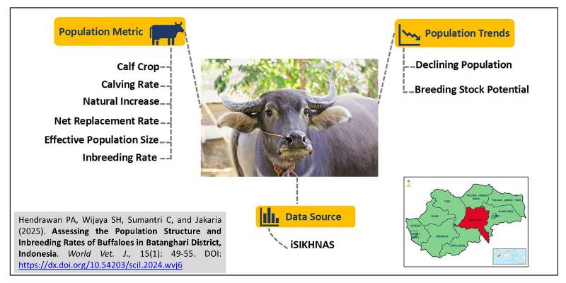
Assessing the Population Structure and Inbreeding Rates of Buffaloes in Batanghari District, Indonesia
Hendrawan PA, Wijaya SH, Sumantri C, and Jakaria.
World Vet. J. 15(1): 49-55, 2025; pii:S232245682500006-15
DOI: https://dx.doi.org/10.54203/scil.2025.wvj6
ABSTRACT: Buffaloes are important in animal husbandry, agriculture, and sociocultural and religious activities in Indonesia. The buffalo population has decreased at the national and regional levels, including in the Batanghari District, Jambi Province, Indonesia. This study analyzed the population structure, effective population size, and inbreeding rate of buffalo populations in the Batanghari District, Jambi, Indonesia, based on secondary data. The data population of 3,149 buffaloes used in this study was sourced from the Integrated National Animal Health System (ISIKHNAS) in the Batanghari District in 2023. The results showed a calf crop of 21.71%, a calving rate of 16.61%, a natural increase of 14.74%, and a net replacement rate of 279.51%. The effective population size was 592 heads, and the inbreeding rate was 0.08%. It can be concluded that the natural increase rate of the buffalo population in the Batanghari District was low, but the number of young replacement animals was sufficient. The effective population size was 592 heads, and the level of inbreeding per generation remained within acceptable limits. Although the buffalo population in the Batanghari District exhibited a negative trend, it still had potential as a source of breeding stock, as indicated by the replacement rate.
Keywords: Buffalo, Effective population, Inbreeding rate, Population structure
[Full text-PDF] [Crossref Metadata] [Scopus] [Export from ePrint]
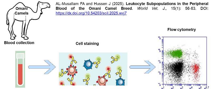
Leukocyte Subpopulations in the Peripheral Blood of the Omani Camel Breed
AL-Musallam FA and Hussen J.
World Vet. J. 15(1): 56-63, 2025; pii:S232245682500007-15
DOI: https://dx.doi.org/10.54203/scil.2025.wvj7
ABSTRACT: Breed-specific variations in immune responses have been studied across various species and breeds. The identification of camel breeds with high immune competence can enhance the breeding of camels with superior immune responsiveness. To date, no study has examined the immune cell composition in the blood of the Omani camel breed. The present study aimed to analyze the immunophenotype of blood leukocytes in the Omani camel breed and investigate the impact of age and gender on the tested immune parameters. To do so, blood samples were collected from 32 clinically healthy camels, randomly selected and comprising 17 camel calves (8 males and 9 females) and 15 adult camels (4 males and 11 females). The samples were tested using flow cytometry and membrane immune fluorescence. The results of the present study revealed a significantly lower count of white blood cells (WBC) in the Omani camel breed than the reference ranges reported for dromedary camels in the published literature. The leukogram was characterized by the dominance of neutrophils (54-60 %) in the blood, followed by lymphocytes (23-33 %). When compared to adult camels, the leukogram pattern in young Omani camels was characterized by elevated WBC and lymphocytes but low levels of eosinophilic granulocytes. The analysis of lymphocyte subsets revealed the dominance of gd T cells over helper T cells and B cells in the blood of young camel calves, confirming that camels belong to the gd T cell-rich species. In addition, lower numbers of B cells and helper T cells in young camels suggest lower cell-mediated and humoral immune functionality compared to adults. Although some differences were identified between male and female adult camels, these results are limited by the low numbers of male camels within the adult group. In conclusion, the distinct leukogram patterns observed in young and adult camels highlight the significant impact of age on the immune competence of Omani camels.
Keywords: Camel, Omani camel, Immunophenotype, Leukocyte
[Full text-PDF] [Crossref Metadata] [Scopus] [Export from ePrint]
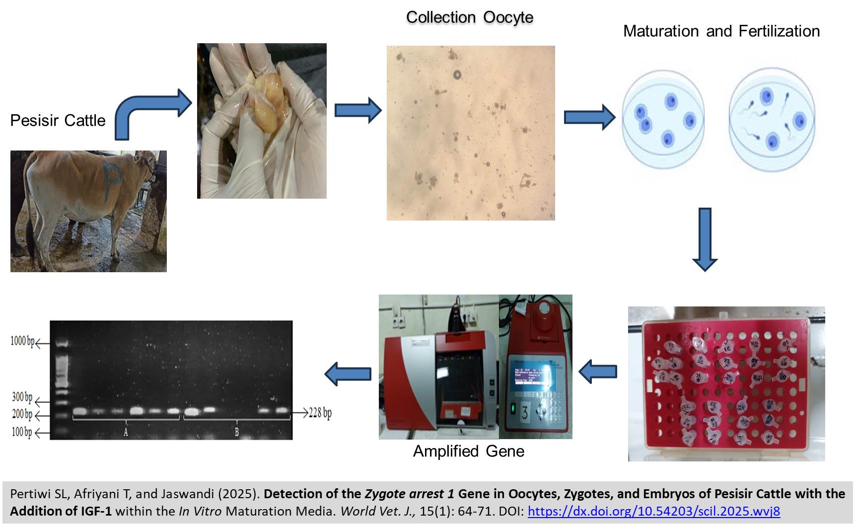
Detection of the Zygote arrest 1 Gene in Oocytes, Zygotes, and Embryos of Pesisir Cattle with the Addition of IGF-1 within the In Vitro Maturation Media
Pertiwi SL, Afriyani T, and Jaswandi.
World Vet. J. 15(1): 64-71, 2025; pii:S232245682500008-15
DOI: https://dx.doi.org/10.54203/scil.2025.wvj8
ABSTRACT: Zygote arrest 1 (ZAR 1) is a maternal gene that plays a crucial role in the oocyte-to-embryo transition. The present study aimed to investigate the presence or absence of the ZAR 1 gene in oocytes, zygotes, and embryos of Pesisir Cattle. Ovaries were collected from cattle at slaughterhouses, and oocytes were retrieved at the Biotechnology Laboratory. The collected oocytes were matured in a maturation medium supplemented with Insulin-like Growth Factor-1 (IGF-1) at a concentration of 10 µg/ml or without IGF-1 for 24 hours in a CO₂ incubator maintained at 38.5°C. Following maturation, the oocytes were fertilized for 18 hours, and the resulting embryos were cultured for 48 hours in a CO₂ incubator at 38.5°C. The samples were then subjected to PCR analysis. The amplification results revealed the presence of the ZAR 1 gene band at the target size of 228 bp in oocytes matured with and without IGF-1. A comparative analysis of oocytes and embryos showed differences in the gene bands, particularly in samples supplemented with IGF-1. These findings suggest that IGF-1 supplementation during oocyte maturation significantly influences ZAR 1 gene expression in embryos. The observed variations in ZAR 1 gene expression across the oocyte, zygote, and embryo stages highlight the gene's pivotal role in reprogramming post-fertilization and maintaining early embryonic development.
Keywords: Embryo, Insulin-like Growth Factor-1, Oocyte, Zygote, Zygote arrest 1 gene
[Full text-PDF] [Crossref Metadata] [Scopus] [Export from ePrint]
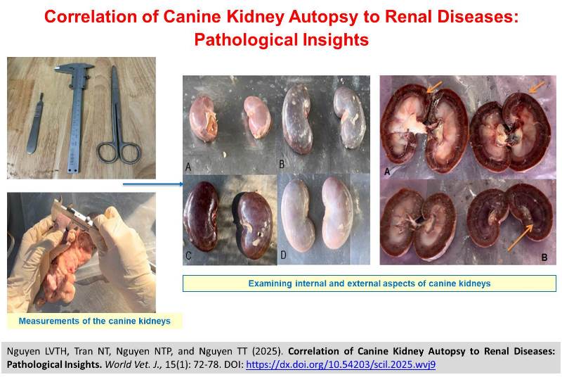
Correlation of Canine Kidney Autopsy to Renal Diseases: Pathological Insights
Nguyen LVTH, Tran NT, Nguyen NTP, and Nguyen TT.
World Vet. J. 15(1): 72-78, 2025; pii:S232245682500009-15
DOI: https://dx.doi.org/10.54203/scil.2025.wvj9
ABSTRACT: Kidneys play a vital role in regulating fluids, electrolytes, hormones, and metabolic waste in canines. This study aimed to enhance the understanding of the correlations between canine kidney autopsy findings and renal diseases. A total of 194 domestic dog samples, including 153 males and 41 females with an average age of 3.88 years (ranging from 1 to 7 years), were physically examined using post-mortem evaluations to understand the prevalence and characteristics of kidney diseases, focusing on both external and internal examinations of the kidneys. Key parameters such as kidney size, texture, and coloration were measured to provide insights into the overall kidney health of the canine population in Vietnam. Results indicated that 22.68% of the dogs had kidney cysts, 29.38% showed signs of external hemorrhage, and 52.06% of the cases exhibited internal hemorrhage, proving to be a condition linked to increased renal vascular resistance and further potentially contributing to renal dysfunction. No evidence of necrosis was detected, and the majority of renal capsules (90.98%) were easy to peel off for further analysis. Kidney size and weight varied obviously in dogs presenting with specific hemorrhagic conditions. This study emphasized the importance of external and internal kidney evaluations in diagnostic measurements and treatment protocols for canine renal diseases while also providing further insights into the current status of the canine population in Vietnam.
Keywords: Dog, Hemorrhage, Kidney, Post-mortem, Renal disease
[Full text-PDF] [Crossref Metadata] [Scopus] [Export from ePrint]
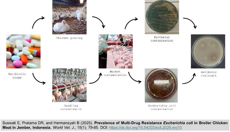
Prevalence of Multi-Drug Resistance Escherichia coli in Broiler Chicken Meat in Jember, Indonesia
Suswati E, Pratama DR, and Hermansyah B.
World Vet. J. 15(1): 79-85, 2025; pii:S232245682500010-15
DOI: https://dx.doi.org/10.54203/scil.2025.wvj10
ABSTRACT: Antibiotic resistance has become one of the global health problems nowadays. Chicken meat is one of the largest food commodities in the world. Escherichia coli (E. coli) is one of the bacteria that is often found in chicken meat. These bacteria are capable of being pathogenic in both animals and humans. This study aimed to determine the prevalence of multidrug-resistant E. coli isolated from broiler chicken meat in the study location. The E. coli utilized in this study were derived from 25 grams of chicken meat obtained from 30 samples procured from six markets within the Jember district. The resistance test method used was Kirby-Bauer with Mueller-Hinton media. The results of the study showed that 100% of chicken meat was contaminated with E. coli. All isolated E. coli from samples in the study were multidrug-resistant. E. coli was 100% resistant to cotrimoxazole and cefixime, 96.67% resistant to chloramphenicol and amoxicillin-clavulanic, 93.3% resistant to tetracycline, 90% resistant to ceftriaxone, and 80% resistant to azithromycin and ciprofloxacin. The minimum resistance profile to 5 types of antibiotics with a multiple antibiotic resistance (MAR) index was between 0.625-1. Thus, the study revealed a high risk of infection associated with the consumption of uncontrolled chicken meat.
Keywords: Antibacterial agent, Chicken, Escherichia coli, Multi-drug resistance
[Full text-PDF] [Crossref Metadata] [Scopus] [Export from ePrint]
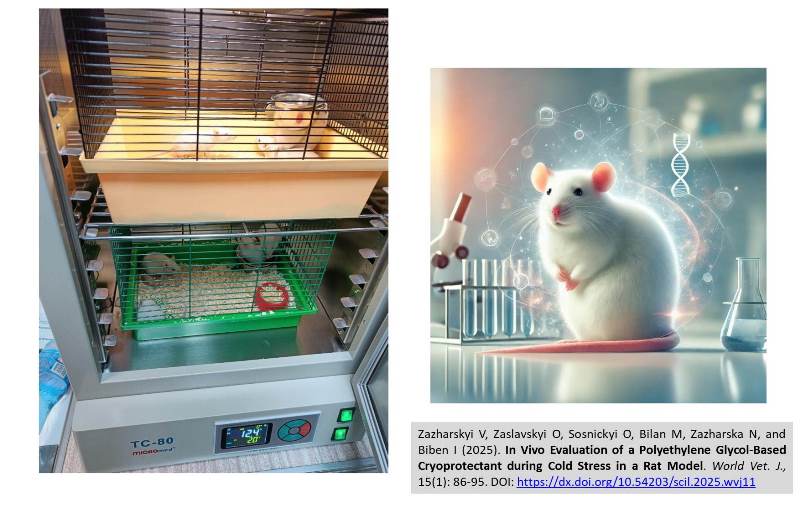
In Vivo Evaluation of a Polyethylene Glycol-Based Cryoprotectant during Cold Stress in a Rat Model
Zazharskyi V, Zaslavskyi O, Sosnickyi O, Bilan M, Zazharska N, and Biben I.
World Vet. J. 15(1): 86-95, 2025; pii:S232245682500011-15
DOI: https://dx.doi.org/10.54203/scil.2025.wvj11
ABSTRACT: Cold stress is an environmental factor that impacts the viability of animals and humans. This study aimed to determine the effectiveness of a cryoprotectant based on polyethylene glycol in reducing cold stress in laboratory rats. For the experiment, 30 outbred Wistar rats (5 weeks) with an average body weight of 55.1 ± 5.3 g were used. Three groups of animals were formed (10 rats per group). The first group served as a positive control, kept at a room temperature of +18 – +20 °С and received 0.1 ml of 0.9% NaCl solution. The second group, the negative control, was kept in critically low temperatures (+2–+4°С) and administered 0.1 ml of 0.9% NaCl solution per experimental animal. Rats of the third group were subjected to cold stress and received 0.1 ml of the experimental preparation. Cold stress in laboratory rats was created using a cooling thermostat. Observation for 14 days included monitoring the dynamics of changes in the live weight of animals (before the start of the experiment, and on days 7 and 14 of the observation) as well as biochemical and haematological blood indicators. Fecal samples were collected from the rectum to determine the qualitative and quantitative state of the intestinal microbiota. The survival level of animals that received the experimental drug within fourteen days was 80.0%, compared to only 40% in the untreated group. When using the experimental drug in laboratory animals, an increase in body weight was noted. The number of full-fledged Escherichia coli in rats that received the drug was 3.4 times higher than the indicator of the group of animals that was kept at a critically low temperature without the drug. The prolonged low temperature in control rats had a negative effect on the animal's body as evidenced by increased leukocyte counts and ALT levels, as well as decreased ALT/AST ratio, and total bilirubin. The use of an experimental polyethylene glycol-based preparation had a positive effect on the weight of rats, blood parameters, and intestinal microbiota of rats under cold stress.
Keywords: Biochemistry, Blood morphology, Cold stress, Cryoprotectant, Intestinal microbiota, Rat
[Full text-PDF] [Crossref Metadata] [Scopus] [Export from ePrint]
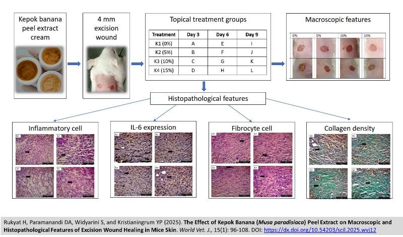
The Effect of Kepok Banana (Musa paradisiaca) Peel Extract on Macroscopic and Histopathological Features of Excision Wound Healing in Mice Skin
Rukyat H, Paramanandi DA, Widyarini S, and Kristianingrum YP.
World Vet. J. 15(1): 96-108, 2025; pii:S232245682500012-15
DOI: https://dx.doi.org/10.54203/scil.2025.wvj12
ABSTRACT: Kepok banana peel extract is known to have a bioactive content that can accelerate wound healing. The present study sought to evaluate the effects of Kepok banana peel extract on the macroscopic and histopathological features of excision wound healing in mouse skin. A total of 24 BALB/c mice were divided into four treatment groups, with each group consisting of six mice. The mice were further divided into three subgroups based on observation days, including days 3, 6, and 9. Each mouse received two excision wounds. The four treatment groups included K1 (control), K2 (topical therapy using Kepok banana peel extract ointment 5%), K3 (topical therapy using Kepok banana peel extract ointment 10%), and K4 (topical therapy using Kepok banana peel extract ointment 15%). The Kepok banana peel extract was obtained using the maceration method, and the ointment Kepok was prepared as a cream with extract concentrations of 5%, 10%, and 15%, using bio cream as the base. Wound healing activity was evaluated across three phases, including inflammatory, proliferative, and remodeling. The parameters observed in the current study included macroscopic and histopathological characteristics of the wound. Macroscopic observations involved wound size, while histopathological analysis included quantification of inflammatory cells, fibrocytes, collagen density, and interleukin-6 expression. Therapy using Kepok banana peel extract ointment was administered for 9 days in the treatment groups. Macroscopic features of the wounds were observed daily, and skin samples from each group were collected on days 3, 6, and 9. The results demonstrated that the 5%, 10%, and 15% concentrations of Kepok banana peel extract formed wound healing areas on mouse skin on days 3, 6, and 9, and were able to reduce the number of inflammatory cells on days 3, 6 and 9 able to reduce IL-6 expression on days 3, 6 and 9, unable to increase fibrocytes on day 3, 6, and 9 and able to increased collagen density on days 6 and 9. The 15% concentration of Kepok banana peel extract applied for 9 days showed the greatest potential to accelerate wound-healing.
Keywords: Histopathological feature, Kepok banana peel, Macroscopic feature, Ointment, Skin, Wound healing
[Full text-PDF] [Crossref Metadata] [Scopus] [Export from ePrint]
Effects of Adding Natural Honey to Semen Extender on Ram Epididymal Sperm Quality
Hasan BJ, Al-Mutar HAH, Taher JK, and Hussain SO.
World Vet. J. 15(1): 109-116, 2025; pii:S232245682500013-15
DOI: https://dx.doi.org/10.54203/scil.2025.wvj13
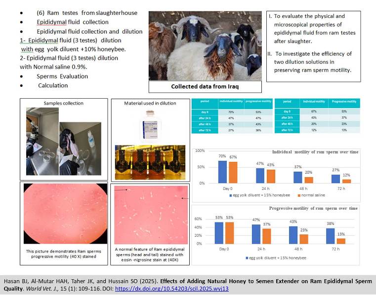
ABSTRACT: Numerous studies have indicated that male infertility is often associated with poor semen quality. The present study investigated the use of natural honey as an antioxidant and nutrient additive to semen extender to evaluate its effect on sperm parameters. A total of 16 fresh testes from sexually mature rams were collected for the investigation and immediately transported to the reproductive technology laboratory at the College of Veterinary Medicine, Baghdad University. For the first group, sperm was collected and diluted with an egg yolk extender mixed with 10% natural honey, while the second group consisted of epididymal sperm diluted with 0.9% normal saline. Epididymal fluid was collected and evaluated for both groups. The results showed significant differences in mean individual motility between the two groups after 48 and 72 hours, as determined by the comparison of proportions. Egg yolk plus honey diluent was significantly more effective than normal saline diluent in preserving sperm cell viability after 48 and 72 hours. The same finding applied to progressive motility; the egg yolk plus honey diluent was significantly more efficient than the normal saline diluent for the time frame after 48 and 72 hours, respectively. In conclusion, the findings demonstrated that the egg yolk extender supplemented with 10% honey was more effective in preserving ram sperm motility over time than normal saline. The addition of honey to the egg yolk extender improved the motility, the live-dead ratio, and the viability of the liquid storage of ram epididymal fluid. Furthermore, egg yolk plays a crucial role in protecting sperm from the detrimental effects of low temperatures.
Keywords: Epididymal sperm, Honey, Ram, Semen extender, Sperm motility
[Full text-PDF] [Crossref Metadata] [Scopus] [Export from ePrint]
Evaluation of Immunomodulatory Properties of Fish-Protein Hydrolysate from Skipjack Tuna by-products (Katsuwonus pelamis, Linnaeus 1758) in Streptozotocin-Nicotinamide-Induced Diabetic Rats
Prasetyo DYB, Agustini TW, Anjani G, and Riyadi PH.
World Vet. J. 15(1): 117-125, 2025; pii:S232245682500014-15
DOI: https://dx.doi.org/10.54203/scil.2025.wvj14
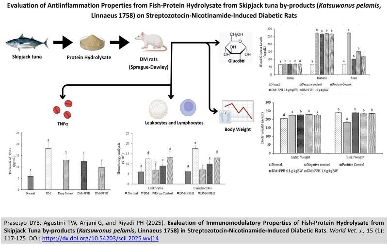
ABSTRACT: Fish protein hydrolysate (FPH) is an alternative to managing fish by-products. Protein hydrolysis by proteolytic enzymes breaks down the protein into bioactive peptides (BP). Bioactive has a high-protein content and various beneficial health properties, including antioxidants, immunoregulatory, antibacterial, anti-inflammatory, and other activities. The current study aimed to investigate the anti-diabetic and immunomodulatory activities of FPH from skipjack tuna by-products. Male Sprague Dawley rats (n = 25) were equally divided into five groups: healthy group, diabetic mellitus (DM) group, DM + Imunos 0.8 g/ kg BW (drug control group), DM + 0.8 g/kg BW (FPH 1), DM + 1.6 g/kg BW FPH (FPH 2). Diabetic rats were induced by being fed with a high-fat diet (HFD) for 3 months, followed by nicotinamide (NA; 120 mg/kg BW)-streptozotocin (STZ) injection (60 mg/kg BW). The initial and final body weights before and after treatment were measured. The leukocyte and lymphocyte levels were measured using a hematology analyzer. The pro-inflammatory cytokine tumor necrosis factor α (TNFα) level was measured using enzyme-linked immunosorbent assay (ELISA). The result showed that the blood glucose levels after treatment using FPH significantly decreased compared with DM rats. Leukocyte and lymphocyte numbers also decreased significantly after treatment using FPH 1 than in DM rats. The pro-inflammatory cytokine TNFα in the FPH rat groups improved significantly compared with DM rats. These study results suggested that FPH from skipjack tuna by-product administration can be used as anti-diabetic and immunomodulatory candidates.
Keywords: Diabetes mellitus, Fish protein hydrolysate, Inflammation, Skipjack tuna by-product
[Full text-PDF] [Crossref Metadata] [Scopus] [Export from ePrint]
Virulent Genes and Genetic Relationship of Salmonella spp. Isolated from Chickens and Husbandry Environments in Small-Scale Farms in the Mekong Delta, Vietnam
Nguyen TK, Huynh LM, Vo TVD, Tran DD, Kha TT, Ly KTL, and Nguyen CTH.
World Vet. J. 15(1): 126-133, 2025; pii:S232245682500015-15
DOI: https://dx.doi.org/10.54203/scil.2025.wvj15
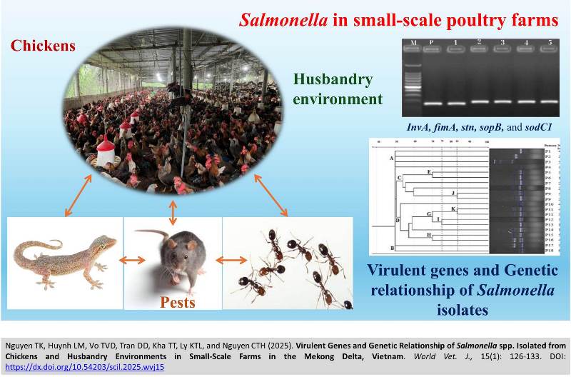
ABSTRACT: Salmonella is one of the most severe pathogens causing diseases in poultry and humans, and several factors could become transmission vectors in the husbandry environment. This study was conducted from April to July 2024 to clarify the prevalence of common Salmonella serovars in chickens and the husbandry environment and their pathogenicity and genetic relationship in small-scale farms in the Mekong Delta, Vietnam. A total of 279 samples were randomly collected from fresh chickens’ feces (n = 54), husbandry environment (n=81), and pests (n=144), including rats, geckos, and ants, in four small-scale farms to examine the prevalence of Salmonella spp. By the conventional isolation method, 75 samples were positive for Salmonella, accounting for 26.88%. The prevalence of Salmonella in chicken feces, the environment, and pests were 27.78%, 12.35%, and 34.72%, respectively. Of 75 positive Salmonella isolates, two common serovars were identified, including S. Gallinarum (13.33%) and S. Enteritidis (10.67%); however, S. Pullorum and S. Typhimurium were not detected using PCR. These Salmonella isolates were detected virulent genes by using PCR, and found that these isolates harbored several virulent genes, including InvA (100%), fimA (100%), stn (93.33%), sopB (89.33%), and sodC1 (54.57%). The ERIC-PCR method was used to determine the genetic relationship among Salmonella strains carrying virulent genes present in chickens, environment, and pests in these small-scale farms. The results showed diversity in phenotype and similarity in the genetic relationship (more than 75% similarity) among Salmonella strains isolated from chicken feces and the livestock environment. In conclusion, the study indicated that pathogenic Salmonella serovars could survive and be transmitted among sources, including chickens, the husbandry environment, and pests in small-scale poultry farms in the Mekong Delta.
Keywords: Chicken, Environment, Genetic relationship, Pest, Salmonella, Virulent gene
[Full text-PDF] [Crossref Metadata] [Scopus] [Export from ePrint]
The Effect of Adding Various Concentrations of Melatonin to Beltsville Thawing Solution Diluent on Berkshire Boar Semen Quality
Oktaviandari PR, Bebas W, Pemayun TGO, Susari NNW, and Laksmi DNDI.
World Vet. J. 15(1): 134-142, 2025; pii:S232245682500016-15
DOI: https://dx.doi.org/10.54203/scil.2025.wvj16
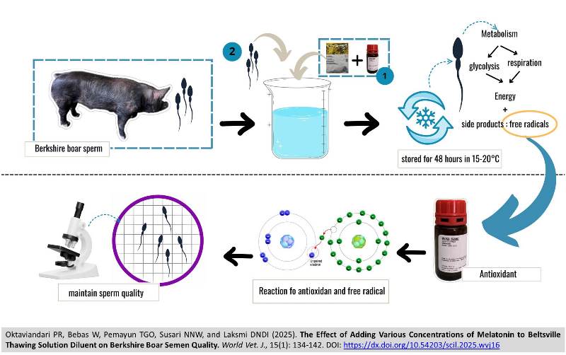
ABSTRACT: Boar spermatozoa contain polyunsaturated fatty acids, rendering them susceptible to damage from free radicals. Oxidative stress in liquid semen can be prevented by modifying the diluent by adding antioxidants. Melatonin is an indoleamine compound that can be used as an antioxidant with high potential in the reproductive system. This study aimed to determine the effect of adding various concentrations of melatonin to Beltsville Thawing Solution (BTS) diluent on the quality of Berkshire boar semen. A Completely Randomized Design (CRD) method was employed, with the experiment divided into four treatment groups, each replicated six times. A total of 24 samples were used in this study. P0 was used as the control group, while groups P1, P2, and P3 were given the addition of melatonin to BTS diluent with different doses, namely 0.5 mM, 1.0 mM, and 1.5 mM. The samples were stored for 48 hours and then examined for boar semen quality. The variables examined included the percentage of motility, abnormality, viability, plasma membrane integrity, and malondialdehyde (MDA) levels. The results showed that the addition of a melatonin dose of 1.0 mM to the BTS diluent was the optimal concentration that could maintain motility, abnormalities, viability, plasma membrane integrity, and MDA levels in Berkshire boar semen compared to other treatment groups and the control group. This study indicated that melatonin functions as an effective antioxidant, neutralizing free radicals and thereby inhibiting oxidative stress in Berkshire boar semen.
Keywords: Beltsville thawing solution, Berkshire boar, Malondialdehyde, Melatonin, Semen quality
[Full text-PDF] [Crossref Metadata] [Scopus] [Export from ePrint]
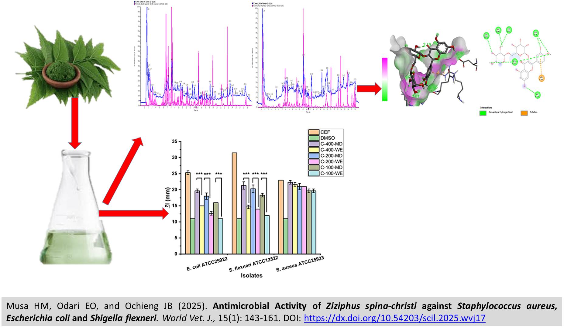
Antimicrobial Activity of Ziziphus spina-christi against Staphylococcus aureus, Escherichia coli and Shigella flexneri
Musa HM, Odari EO, and Ochieng JB.
World Vet. J. 15(1): 143-161, 2025; pii:S232245682500017-15
DOI: https://dx.doi.org/10.54203/scil.2025.wvj17
ABSTRACT: Antibiotic resistance remains a global concern, with up to 1.91 million deaths projected to occur due to resistant Gram-negative and Gram-positive bacteria by 2050. The study hence aimed to assess the antimicrobial activity of Ziziphus spina-christi leaf extracts in relation to specific bacterial strains and elucidate the molecular mechanisms to validate the in vitro findings. Ziziphus spina-christi leaf extracts were tested against Staphylococcus aureus (S. aureus), Escherichia coli (E. coli), and Shigella flexneri (S. flexneri). The leaf powder was subjected to both aqueous and methanol-dichloromethane extraction. Phytochemical products were determined by Liquid Chromatography-Mass Spectrometry and Gas Chromatography-Mass Spectrometry for water extract and methanol dichloromethane extract, respectively. The agar well diffusion method, broth microdilution, and minimum bactericidal concentration against three bacterial species, S. aureus, E. coli, and S. flexneri, were used to assess the antibacterial activity of extracts. The results have shown that both plant extract has a significant level of antibacterial activity at higher concentrations (400 mg/ml) against the gram-positive bacteria. In addition, the methanol-dichloromethane extract exhibited the highest antibacterial activity against Gram-negative bacteria (S. flexneri, and E. coli), conversely, the water extract demonstrated a lower activity against S. flexneri and E. coli, with inhibition zones of 15 ± 0 mm for both bacteria. At a lower concentration (100 mg/ml), the methanol-dichloromethane extract produced inhibition zones of 19.6 ± 0.5 mm against S. aureus, closely followed by S. flexneri and E. coli. The water extract exhibited high antibacterial activity against Gram-positive bacteria. However, exhibited reduced antibacterial activity against S. flexneri and E. coli, indicating a concentration-dependent antibacterial effect. Extraction methods were significantly different, with products generated from non-aqueous extraction demonstrating a higher potency against both Gram-negative and Gram-positive bacteria than the aqueous extract. Docking results demonstrated that water extract had a high binding activity against penicillin-binding proteins. Moreover, it serves as a potent beta-lactamase inhibitor as it binds to their active site, rendering them inactive and inhibiting the hydrolysis of Beta lactam antibiotics. In conclusion, the methanol-dichloromethane and water Ziziphus spina-christi leaves could be considered a promising source of antimicrobial ingredients.
Keywords: Antimicrobial resistance, Beta lactamase, Methanol-dichloromethane Extract, Molecular docking, Ziziphus spina-christi leave
[Full text-PDF] [Crossref Metadata] [Scopus] [Export from ePrint]
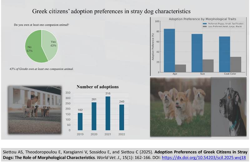
Adoption Preferences of Greek Citizens in Stray Dogs: The Role of Morphological Characteristics
Siettou AS, Theodoropoulou E, Karagianni V, Sossidou E, and Siettou C.
World Vet. J. 15(1): 162-166, 2025; pii:S232245682500018-15
DOI: https://dx.doi.org/10.54203/scil.2025.wvj18
ABSTRACT: A critical aspect of addressing the stray dog population issue in Greece involves the investigation of the likelihood of dog adoptions based on morphological traits. Recent statistics indicate that 43% of Greek households own at least one companion animal. This study aimed to explore some of the factors influencing the decision to adopt a stray and the morphological traits that are preferred by the adopters. To undertake this research, data spanning four years (2019-2022) were used; the data comprised information on 858 adoptions from a Greek animal welfare organization on the Island of Lesvos in the Aegean Sea. The analysis revealed that younger age, smaller size, and tan or tricolor coats were associated with faster adoption rates. In addition, when comparing the data of all four years (2019-2022), the year of adoption was found to be a statistically significant variable, which confirms that Greeks also followed the international trend of increased adoptions during the Covid-19 pandemic.
Keywords: Adopter preference, Greece, Morphological trait, Shelter dog, Stray companion animal
[Full text-PDF] [Crossref Metadata] [Scopus] [Export from ePrint]
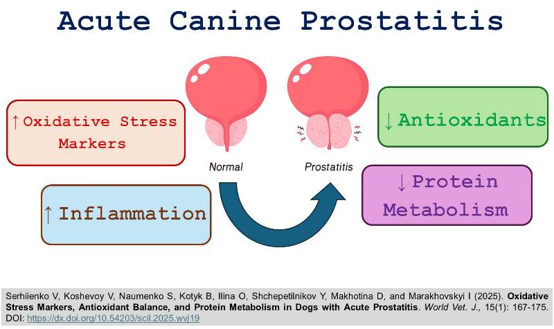
Oxidative Stress Markers, Antioxidant Balance, and Protein Metabolism in Dogs with Acute Prostatitis
Serhiienko V, Koshevoy V, Naumenko S, Kotyk B, Ilina O, Shchepetilnikov Y, Makhotina D, and Marakhovskyi I.
World Vet. J. 15(1): 167-175, 2025; pii:S232245682500019-15
DOI: https://dx.doi.org/10.54203/scil.2025.wvj19
ABSTRACT: The prostate gland in dogs is highly vulnerable to the action of negative pathogens due to its structure and topography. Among the numerous etiological factors in the development of prostatitis, inflammatory processes and oxidative stress play a predominant role, regardless of whether the condition is bacterial, viral, or autoimmune in origin. This study aimed to assess protein metabolism and redox balance indicators in the prostate tissue of dogs with acute prostatitis. For biochemical analyses, prostate tissue samples were taken from 24 mixed-breed dogs, including twelve animals that were considered healthy with no abnormalities of the genitourinary system (control group) and twelve animals with newly diagnosed acute prostatitis, from which samples were obtained via biopsy (experimental group). Following homogenization and sample preparation, all biochemical parameters in the prostate tissue were determined spectrophotometrically. The results of biochemical studies in dogs with acute prostatitis demonstrated a significant increase in the content of thiobarbiturate acid-reactive compounds by 102.2% and the level of lipid hydroperoxides by 35.7% compared to healthy dogs in the control group. In contrast, the total protein content was 32.9% lower than in the control group, while reduced glutathione levels decreased by 76.5%. Similar changes to the dynamics of oxidative stress markers were indicated by the activity of antioxidant enzymes, with glutathione peroxidase and catalase activities increasing by 61.3% and 21.8%, respectively, relative to the control group. These findings indicate the presence of oxidative stress in dogs with acute prostatitis. The biochemical changes observed in prostate tissue provide a foundation for future research aimed at developing therapeutic methods that incorporate anti-inflammatory, antibacterial, and antioxidant agents for the treatment of acute prostatitis in dogs.
Keywords: Biochemistry, Dog, Inflammation, Oxidative stress, Prostate
[Full text-PDF] [Crossref Metadata] [Scopus] [Export from ePrint]
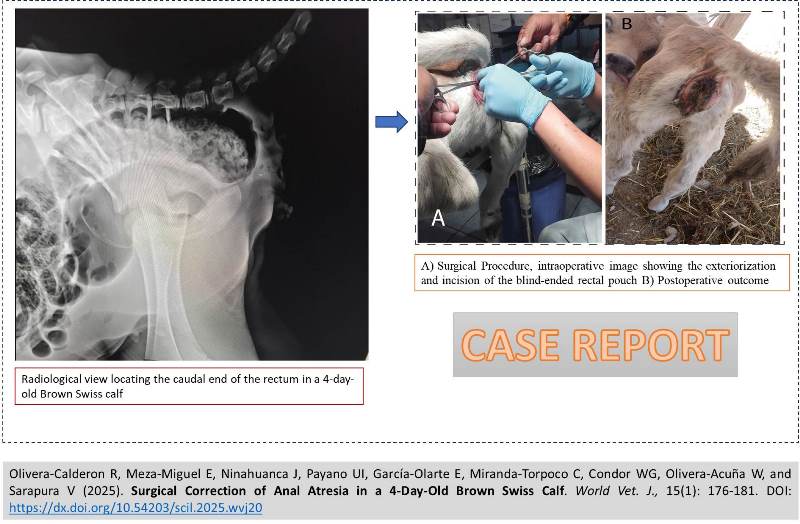
Surgical Correction of Anal Atresia in a 4-Day-Old Brown Swiss Calf
Olivera-Calderon R, Meza-Miguel E, Ninahuanca J, Payano UI, García-Olarte E, Miranda-Torpoco C, Condor WG, Olivera-Acuña W, and Sarapura V.
World Vet. J. 15(1): 176-181, 2025; pii:S232245682500020-15
DOI: https://dx.doi.org/10.54203/scil.2025.wvj20
ABSTRACT: A 4-day-old Brown Swiss calf was presented to a veterinary clinic in Huancayo, Peru, with congenital anal atresia. The condition was characterized by an absence of defecation, progressive abdominal distension, and a blind rectal pouch confirmed radiographically. Clinical examination revealed no anal opening, moderate tympany, and normal neurological function. Surgical correction was conducted under pre-anesthetic sedation using xylazine (0.2 mg/kg IM) and sacrococcygeal epidural anesthesia with lidocaine (0.5 mL/kg). A 12-cm vertical incision in the intertubercular ischiatic region exposed the distended rectal pouch, allowing for an end-to-cutaneous anastomosis with polyglactin 910 (Vicryl® 2-0). Subsequently, a 2-3 cm distal rectal incision was created to form a neoanus. The mucosal edges were sutured to the skin margins with interrupted simple stitches (Vicryl® 3-0), a technique aimed at preventing stenosis and maintaining a neoanal diameter of approximately 1.5-2 cm. Postoperative management included prophylactic penicillin. Within three hours of surgery, the calf passed impacted meconium, indicating the functional patency of the neoanus. Although the prognosis was favorable, the patient was closely monitored for five days to detect any signs of stricture or infection. This case illustrated the effectiveness of early surgical intervention in reestablishing gastrointestinal continuity in congenital anal atresia.
Keywords: Anal agenesis, Clinical significance, Postoperative outcome, Surgical technique
[Full text-PDF] [Crossref Metadata] [Scopus] [Export from ePrint]
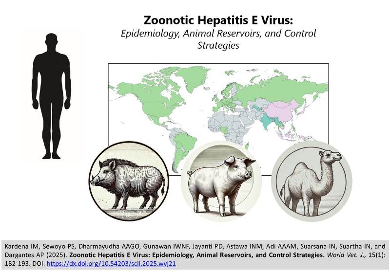
Zoonotic Hepatitis E Virus: Epidemiology, Animal Reservoirs, and Control Strategies
Kardena IM, Sewoyo PS, Dharmayudha AAGO, Gunawan IWNF, Jayanti PD, Astawa INM, Adi AAAM, Suarsana IN, Suartha IN, and Dargantes AP.
World Vet. J. 15(1): 182-193, 2025; pii:S232245682500021-15
DOI: https://dx.doi.org/10.54203/scil.2025.wvj21
ABSTRACT: Hepatitis E virus (HEV) is a leading cause of acute hepatitis transmitted through the enteric route, impacting millions of individuals worldwide annually. While HEV is generally self-limiting, it can lead to considerable illness and death, particularly in gestating women, immunocompromised individuals, and those with pre-existing conditions, such as chronic liver disease. Hepatitis E virus genotypes 1 and 2 infect solely humans and are prevalent in areas with poor sanitation, whereas genotypes 3 and 4 are zoonotic, infecting both animals and humans. Hepatitis E virus genotype 7 has been reported to infect both humans and animals; however, further research is needed to clarify its zoonotic potential. Zoonotic transmission occurs primarily through the consumption of contaminated meat and close contact with infected animals, posing significant public health risks. Epidemiological studies indicated an increasing seroprevalence of HEV in humans and animals across diverse regions, highlighting the need for strengthened public health measures. While HEV infections in animal reservoirs are generally asymptomatic, they represent a critical source of human infections. The present review aimed to highlight HEV's current classification, epidemiology, modes of transmission from animals to humans, prevention, and control measures, with a special focus on HEV zoonotic genotypes and their animal reservoirs.
Keywords: Control measure, Epidemiology, Hepatitis E virus, Public health, Zoonotic genotype
[Full text-PDF] [Crossref Metadata] [Scopus] [Export from ePrint]
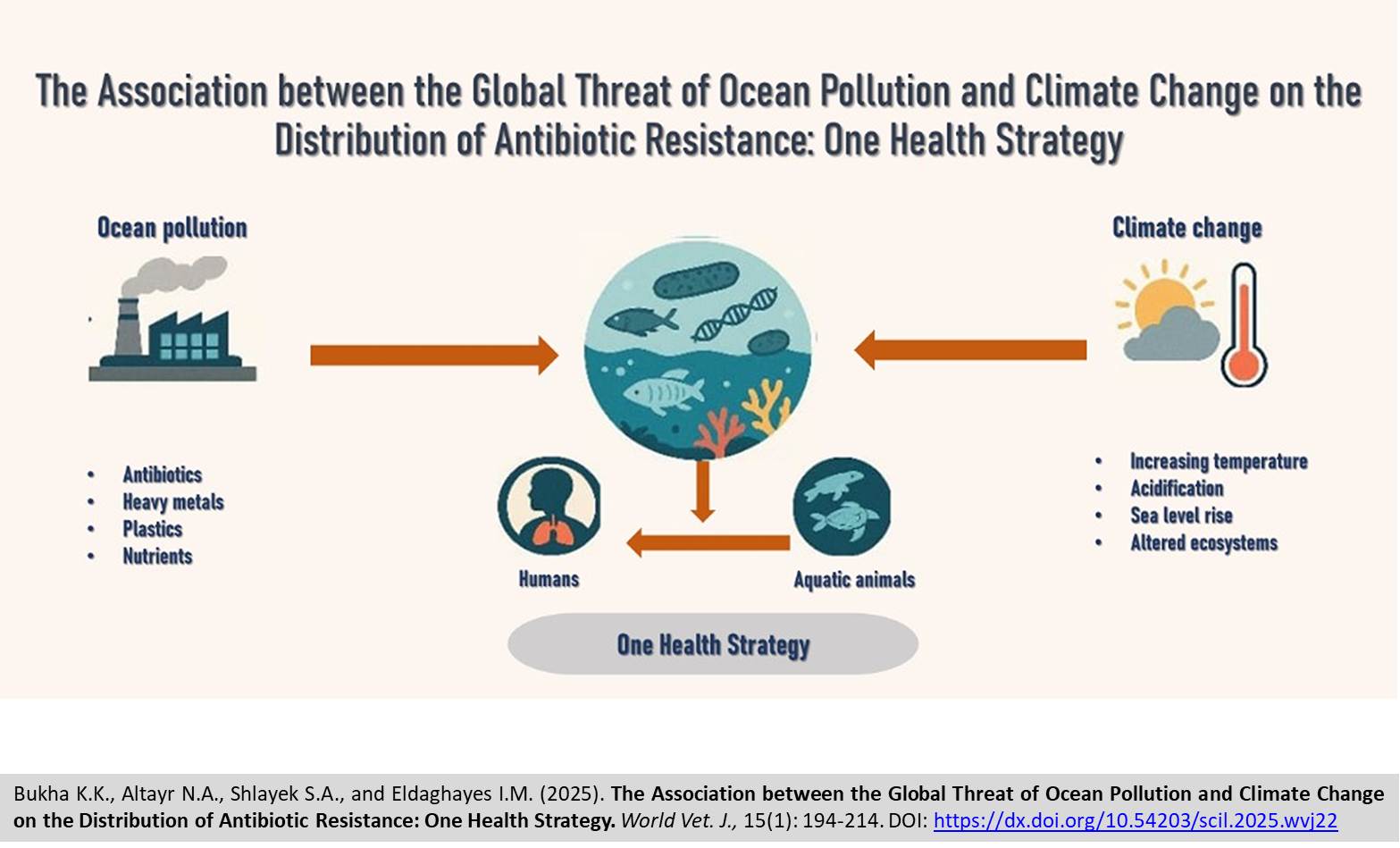
The Association between the Global Threat of Ocean Pollution and Climate Change on the Distribution of Antibiotic Resistance: One Health Strategy
Bukha KK, Altayr NA, Shlayek SA, and Eldaghayes IM.
World Vet. J. 15(1): 194-214, 2025; pii:S232245682500022-15
DOI: https://dx.doi.org/10.54203/scil.2025.wvj22
ABSTRACT: Antibiotic resistance represents a fundamental issue affecting public health today. Antibiotic resistance occurs when bacteria alter their response to antibiotics. Pathogenic bacteria and their genes can diffuse throughout human and natural habitats. The complicated interactions between diverse bacterial communities that affect the health of people, aquatic animals, and the aquatic environment are an illustration of ecological issues. Pollution of the oceans by antibiotic-resistant bacteria (ARB) can disturb the natural equilibrium of the oceans and may spread to humans. Also, climate change (CC) significantly affects the health of marine environments. Rising temperatures, acidification, increased sea levels, an increasing number of invasive marine animals, changed biological systems, and a decrease in biodiversity are some of the major impacts on the oceans caused by CC. However, the elevated temperatures linked to CC facilitate the higher spread of bacterial infections in aquatic environments, aquatic animals, and humans with the emergence of antibiotic resistance. The present study aimed to provide a scientific understanding of the relationship between ocean pollution and CC, as well as their impacts on ocean health. Additionally, the present study presented the current status of ARB and its associated genes in the oceans, comparing to future projections based on previous studies. One Health (OH) concept strategies for reducing antibiotic pollution in the ocean were discussed. The present paper is a foundation for further studies to determine the prevalence of antibiotic resistance in the oceans, as well as to understand the current state and key highlights of ocean pollution.
Keywords: Climate change, Drug-resistant bacteria, Heavy metal resistance gene, Ocean pollution, One Health concept
[Full text-PDF] [Crossref Metadata] [Scopus] [Export from ePrint]
Previous issue | Next issue | Archive
![]() This work is licensed under a Creative Commons Attribution 4.0 International License (CC BY 4.0).
This work is licensed under a Creative Commons Attribution 4.0 International License (CC BY 4.0).


